
Human Cortical Neurons
Cat.No.: CSC-C1514
Species: Human
Source: Brain
Cell Type: Neuron
- Specification
- Background
- Scientific Data
- Q & A
- Customer Review
Human Cortical Neurons (HCN) are terminally differentiated neurons that are not immortalized. They are normally obtained from ethically acquired fetal brain tissues from 14-22 gestational weeks. Unlike immortalized cell lines like SH-SY5Y, these cells maintain the phenotypic, molecular, and electrophysiological characteristics of human cortical neurons in vivo. They express key neuronal markers like TUJ1, MAP2, NeuN, and subtype-specific markers for glutamatergic and GABAergic neurons. They build functioning synaptic networks and have mature electrical activity, giving them an extremely physiologically realistic model. HCN are commonly utilized in neurodevelopment, neurodegenerative disease modeling, and CNS drug screening, and have improved clinical translatability than rodent models, despite constraints like as short culture lifetime, donor variability, and expensive cost.
SARS-CoV-2 S1 Protein Induces Endolysosome Dysfunction and Neuritic Dystrophy
Neurological disorders are increasingly associated with COVID-19, yet their pathogenesis remains unclear due to low or undetectable SARS-CoV-2 levels in brain tissue. The spike S1 protein, which can cross the blood-brain barrier and has been detected in neurons, presents a potential alternative mechanism. Datta et al. aimed to test the hypothesis that SARS-CoV-2 S1 protein directly induces neuronal injury.
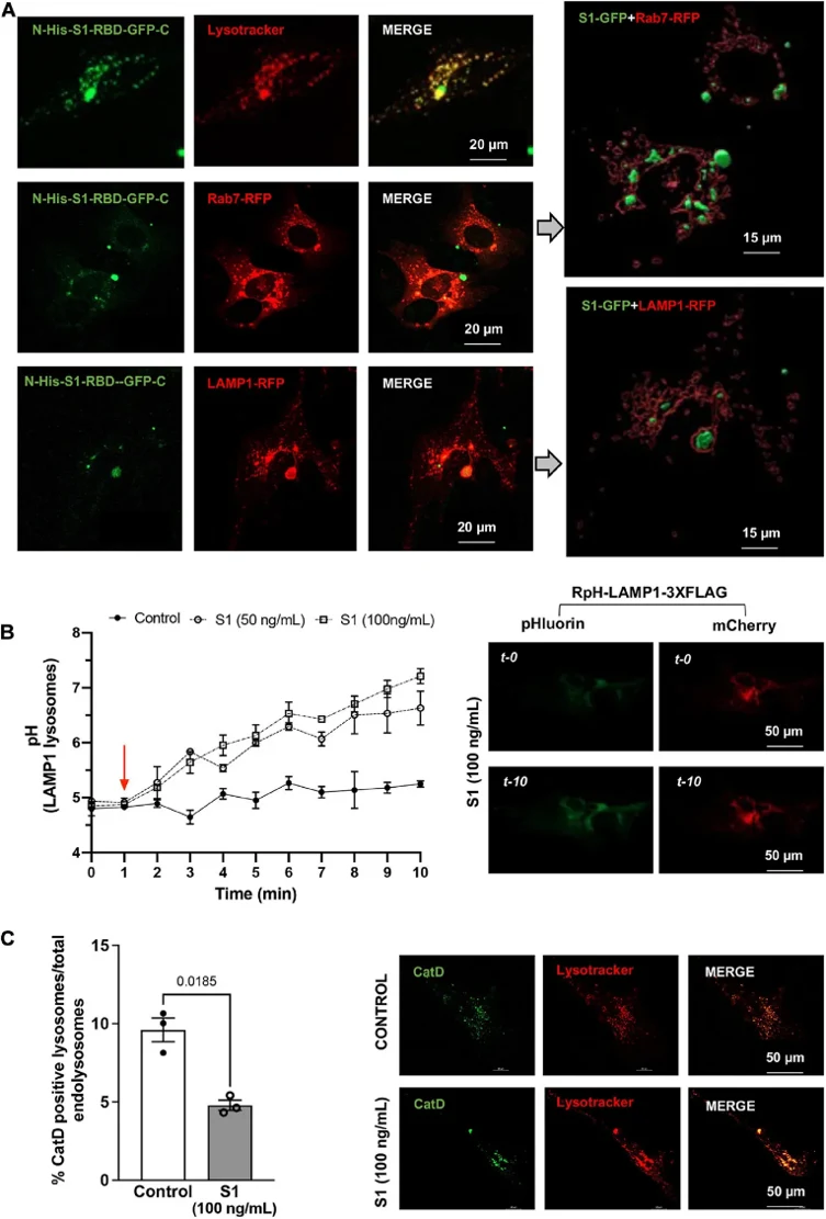
Using primary human cortical neurons, researchers investigated whether SARS-CoV-2 S1 protein accumulation causes cellular damage underlying COVID-19 neurological complications. Following characterization in CLU199 neuronal cells, we examined S1 effects on endolysosome function in primary human cortical neurons. S1-RBD-GFP (250 ng/ml, 48 h) localized to Lysotracker-, Rab7-, and LAMP1-positive endolysosomes (Fig. 1A). S1 protein (50-100 ng/ml) rapidly and robustly de-acidified endolysosomes, measured by ratiometric pH sensing in LAMP1-positive vesicles (Fig. 1B). Additionally, S1 (100 ng/ml, 30 min) decreased the percentage of endolysosomes containing enzymatically active cathepsin D (Fig. 1C), consistent with findings in CLU199 cells.
Ask a Question
Write your own review

Description: Creative Bioarray's Human Monocyte-Derived Dendritic Cells provide an ideal culture model for the study of the initiation of immune responses, delivery of antigens to dendritic cells, cytokine ...

Description: GFP Expressing Human Astrocytes (GFP-HAs) provided by Creative Bioarray are puromycin-selected from primary human astrocytes infected with lentivirus expressing GFP. GFP-HAs are negative for ...

Description: Human Neurons-hippocampal (HN-h) from Creative Bioarray are isolated from hippocampal tissue of the brain. HHN are cryopreserved at primary cultures and delivered frozen. Each vial contains >1 x 10^6 ...
Description: Human Astrocytes-midbrain (HA-mb) from Creative Bioarray are isolated from human midbrain (mesencephalon). HA-mb are cryopreserved at passage one and delivered frozen. Each vial contains >5 x 10^5 ...
Description: Human Choroid Plexus Fibroblasts (HCPF) from Creative Bioarray are isolated from human choroid pluxus. HCPF are cryopreserved at primary culture and delivered frozen. Each vial contains >5 x 10^5 ...
Description: HN are isolated from the human brain. HN are cryopreserved at primary cultures and delivered frozen. Each vial contains >1 x 10^6 cells in 1 ml volume. HN are characterized by immunofluorescent ...