
Human Bronchial Epithelial Cells-COPD
Cat.No.: CSC-C8075L
Species: Human
Source: Bronchus
Cell Type: Epithelial Cell
- Specification
- Background
- Scientific Data
- Q & A
- Customer Review
Human Bronchial Epithelial Cells-COPD (HBEC-COPD) are primary airway epithelial cells obtained from bronchial tissue from donors diagnosed with chronic obstructive pulmonary disease (COPD). These cells exhibit many of the phenotypic and genetic traits associated with illness seen in COPD patients, and hence represent a highly useful in vitro model to research airway pathology and therapeutic approaches. Compared to normal bronchial epithelial cells, COPD-derived HBECs are generally characterized by defective epithelial barrier integrity, aberrant mucociliary differentiation, increased oxidative stress responses, elevated inflammatory signaling, and altered tissue repair ability.
Human Bronchial Epithelial Cells-COPD are frequently utilized to study critical mechanisms of COPD progression including chronic airway inflammation, epithelial remodeling, mucus hypersecretion, ciliary dysfunction and responses to cigarette smoke exposure. These cells can develop into pseudostratified airway epithelium mimicking the in vivo bronchial mucosa when cultivated under air-liquid interface (ALI) conditions, providing a physiologically realistic platform for disease modeling. Apart from COPD research, HBEC-COPD cells are useful models to study respiratory virus infections, host-pathogen interactions, drug screening, toxicology testing and personalized medicine techniques. Their capacity to recreate patient-specific aspects of the disease makes them particularly relevant for the evaluation of innovative anti-inflammatory, anti-fibrotic and regenerative medicines for chronic airway diseases.
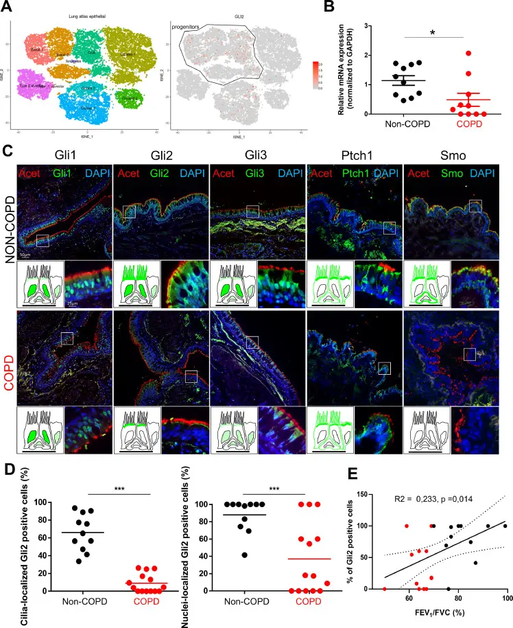
Defective Hedgehog Signaling Localization Correlates with COPD Severity
Chronic obstructive pulmonary disease (COPD) involves pathological airway remodeling. Belgacemi et al. investigated the Hedgehog (HH) pathway, essential for lung development, in human airway epithelial cell (AEC) differentiation and COPD pathology.
Analysis of the Human Lung Atlas showed GLI1-3, SMO, and PTCH1 transcripts localized to progenitor cells (Fig. 1A). In AECs from large airway brushings, COPD patients exhibited a 2.3-fold decrease in GLI2 expression and undetectable SMO compared to non-COPD controls (Fig. 1B). Immunohistochemistry of lung tissues (20 non-COPD, 20 COPD) revealed severe disruption of protein localization in COPD. Non-COPD epithelia showed widespread ciliary and nuclear Gli2, whereas COPD samples had drastically reduced ciliary Gli2 (8.9% vs. 66%) and nuclear Gli2 (37.1% vs. 87.8%) (Fig. 1C, D). Smo localization was similarly impaired, with ciliary presence dropping to 5.78% (vs. 59.9%) and membrane-bound Smo to 32.1% (vs. 98%) (Fig. 1C).
Nuclear Gli2 translocation correlated with disease status. COPD patients had significantly fewer nuclear Gli2-positive cells (37% vs. 77%). A 25% nuclear Gli2 threshold discriminated COPD from non-COPD (AUC = 0.686, OR = 12.833, p= 0.015). Crucially, the percentage of nuclear Gli2-positive cells positively correlated with lung function (FEV₁/FVC) (Fig. 1E). Smo localization showed a similar correlation. These findings indicate that defective HH signaling localization is a hallmark of COPD airway pathology directly linked to disease severity.
Ask a Question
Write your own review
- You May Also Need
Description: Human Bronchial/Tracheal Epithelial Cells-Cystic Fibrosis (HBECs-CF) are isolated from cystic fibrosis human lung tissue. The HSAECs-CF are guaranteed through 15 population doublings.
Description: Human Bronchial Epithelial Cells are isolated from normal human bronchial tissue.
Description: Cryopreserved Diseased Bronchial Smooth Muscle cells stain negative for von Willebrand factor VIII, cytokeratins 18 and 19, and alpha smooth muscle actin. All cells test negative for mycoplasma, ...
Description: The most abundant cell type in the bronchus is fibroblasts. They resemble a mesenchymal stem cell phenotype and their principle function is the production of type III collagen, elastin, and ...
Description: Human hypertension tracheal epithelial cells are isolated from the tracheal tissues of human donor that has been diagnosed with Hypertension. Human Hypertension Tracheal Epithelial Cells are grown in ...
Description: Cryopreserved Bronchial Smooth Muscle cells stain negative for von Willebrand factor VIII, cytokeratins 18 and 19, and alpha smooth muscle actin. All cells test negative for mycoplasma, bacteria, ...