
Human Bladder Smooth Muscle Cells (HBdSMC)
Cat.No.: CSC-7829W
Species: Human
Source: Bladder
Cell Type: Smooth Muscle Cell
- Specification
- Background
- Scientific Data
- Q & A
- Customer Review
Human Bladder smooth muscle cells (HBdSMC) are primary smooth muscle cells cultured from the detrusor layer of the human urinary bladder. Detrusor smooth muscle is the smooth muscle of the bladder wall and is responsible for bladder contractility. Detrusor muscle plays a central role in urine storage and voiding, as it must coordinate relaxation and contraction in response to neural and biochemical cues. HBdSMCs recapitulate the structure, molecular and functional properties of native bladder smooth muscle in vitro.
HBdSMCs exhibit the characteristic spindle-shaped morphology of smooth muscle cells and express canonical smooth muscle markers such as α-smooth muscle actin (α-SMA), smooth muscle myosin heavy chain (SM-MHC), calponin, and desmin. HBdSMCs are responsive to neurotransmitters, growth factors, and inflammatory mediators and can exhibit contractile and proliferative phenotypes in culture, reflecting the physiological and pathological plasticity of bladder smooth muscle in vivo. These cells have been widely used to study bladder development, detrusor contractility, and smooth muscle remodeling in the context of urological disease and injury. HBdSMCs are used as an in vitro model for the study of bladder dysfunctions such as overactive bladder, bladder outlet obstruction, and neurogenic bladder, as well as for drug screening, toxicology, and regenerative therapies targeting bladder smooth muscle.
pSMC-CM Upregulated Extracellular Matrix Elastin Metabolism in Human Bladder Smooth Muscle Cells (bSMCs) and Vaginal Fibroblasts
Adult mesenchymal stem cells (MSCs) have limited proliferation and can induce senescence with prolonged culture. Zhuang et al. investigated the paracrine effects of human smooth muscle cell progenitors (pSMCs) derived from pluripotent stem cells (PSCs) on stress urinary incontinence (SUI) in rodents, aiming to develop a novel therapeutic approach.
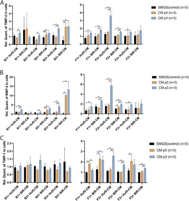
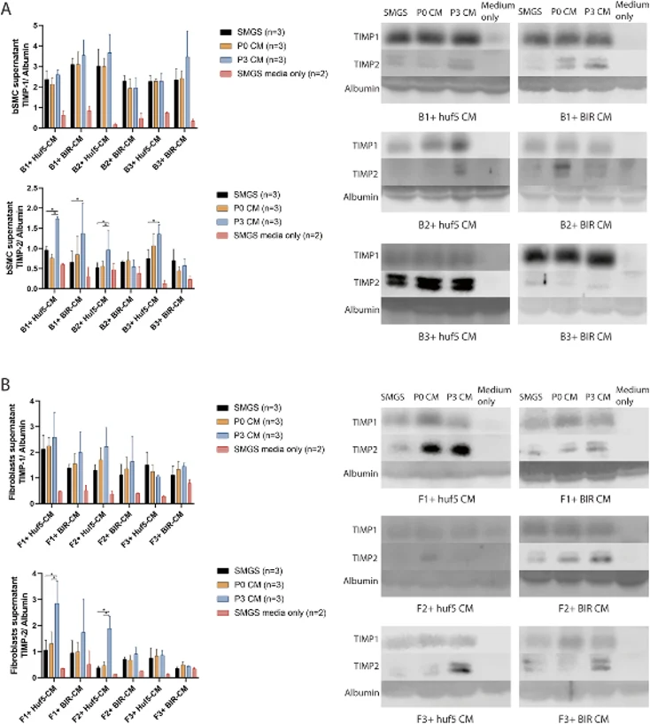
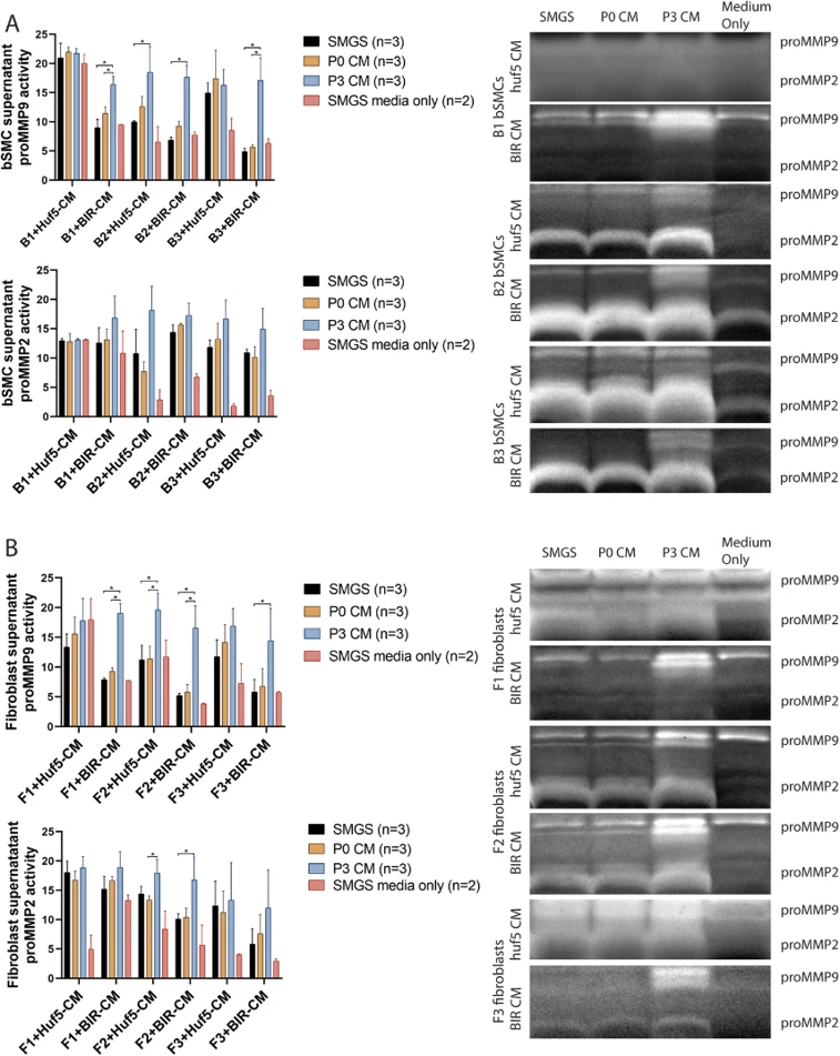
To investigate how pSMC-CM regulates ECM metabolism in human bladder smooth muscle cells (B1, B2, B3) and vaginal fibroblasts (F1, F2, F3), qPCR was performed after treatment with pSMC-CM derived from two human PSC lines (Huf5 and BIR). Compared to control medium, 9 out of 12 groups treated with pSMC-CM showed a significant increase in TIMP-2 and MMP-2 mRNA expression (Fig. 1a, b). TIMP-1 mRNA expression was not significantly different in bSMCs treated with pSMC-CM compared to control medium, but was significantly increased in 4 out of 6 fibroblast groups treated with P3-pSMC-CM (Fig. 1c). Western blot analysis showed that in 4 out of 6 bSMC groups and 2 out of 6 fibroblast groups, cells treated with P3 pSMC-CM secreted more TIMP-2 than those treated with control medium (Fig. 2a, b). Neither bSMCs nor fibroblasts secreted TIMP-1 when cultured with passage 0 or passage 3 pSMC-CM. Nonreducing gelatin zymography revealed that in 8 out of 12 groups, passage 3 pSMC-CM significantly upregulated pro-MMP-9 activity in the supernatant of treated cells (Fig. 3). Pro-MMP-2 activity was similar to controls in all groups.
Yes.
The human smooth muscle bladder cells are sold at passage 2, 3 or 4. Passage 9 is the maximum recommended expansion of the cells.
Average doubling time ranges from 24-48 hours. However, keep in mind that the replication rate varies slightly from donor to donor.
Yes. Penicillin ,streptomycin and amphotericin B are included in the media.
Ask a Question
Average Rating: 5.0 | 1 Scientist has reviewed this product
Wonderful
We have always trusted Creative Bioarray's cell products to contribute to our research.
23 July 2022
Ease of use
After sales services
Value for money
Write your own review
Description: Special edition cells are isolated from the tissue types described. Cells are sterility and virus tested. All tissues used for cell isolation are obtained under informed consent. Creative Bioarray ...

Description: Creative Bioarray's normal Human Bladder Epithelial Cells - Apex, when grown in LIUro A culture medium, provide an ideal culture model for the study of bladder biology. Cell Features:Human Bladder ...

Description: Human Bladder Microvascular Endothelial Cells from Creative Bioarray are isolated from human bladder tissue. Human Bladder Microvascular Endothelial Cells are grown in T25 tissue culture flasks ...
Description: Human Bladder Epithelial Cells are isolated from normal human bladder tissue.
Description: Creative Bioarray's normal Human Bladder Epithelial Cells - Dome, when grown in LIUro D Complete culture medium, provide an ideal culture model for the study of bladder biology.
Description: HBdSF from Creative Bioarray are isolated from human bladder tissue. HBdSF are cryopreserved at secondary culture (passage one) and are delivered frozen. Each vial contains >5 x 10^5 cells in 1 ml ...