
Human Bladder Epithelial Cells - Apex
Cat.No.: CSC-C4104X
Species: Human
Source: Bladder
Cell Type: Epithelial Cell
- Specification
- Background
- Scientific Data
- Q & A
- Customer Review
Cell Features:
Human Bladder Epithelial Cells –Dome are cryopreserved as secondary cells, e.g. cells are isolated from the dome region of human bladder tissue and expanded in culture vessels twice before being harvested for cryopreservation.
Human Bladder Epithelial Cells –Apex are cryopreserved as secondary cells, e.g. cells are isolated from the apex/neck/trigone region of human bladder tissue and expanded in culture vessels twice before being harvested for cryopreservation.
Bladder cells are not exposed to phenol red or antimicrobials when cultured in Creative Bioarray medium.
Creative Bioarray guarantees performance and quality.
Human Bladder Epithelial Cells - Apex are primary urothelial cells isolated from the apical (luminal) layer of the human bladder epithelium. The bladder epithelium, or urothelium, is a specialized stratified epithelium that creates a tight, impermeable barrier to protect underlying tissue from urine toxicity while allowing finely-tuned permeability and stretch in response to bladder filling and voiding. Cells from the apical layer, which are sometimes called umbrella cells, are critical for formation of this impermeable bladder epithelial barrier.
Human Bladder Epithelial Cells show typical epithelial morphology and express urothelial differentiation markers including uroplakins (UPK1A, UPK2, UPK3A), cytokeratins (CK7, CK8, CK18), E-cadherin, and tight junction proteins such as ZO-1 and claudins. Human Bladder Epithelial Cells - Apex show robust barrier properties and high responsiveness to mechanical stretch, inflammatory stimuli, and chemicals, and recapitulate many in vivo physiological properties of bladder urothelial cells. These cells are commonly used in studies of bladder physiology and urothelial differentiation, bladder host-pathogen interactions, inflammatory conditions such as interstitial cystitis, as well as drug permeability, cytotoxicity, and safety of intravesical therapeutics. They are also used in studies of mechanisms of bladder cancer initiation and urothelial dysfunction.
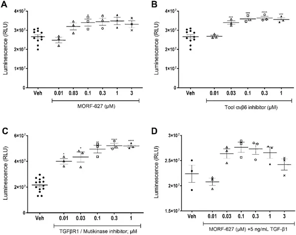
MORF-627-Induced Epithelial Proliferation in Cultured Primary Human Bladder Cells, and Proliferative Changes were Sensitive to TGF-β
Integrin αvβ6 inhibition is intended to block TGF-β signaling, implicated in epithelial proliferative disorders. Guffroy et al. investigated the effects of a selective integrin αvβ6 inhibitor, MORF-627, on urinary bladder epithelium in young monkeys and human bladder epithelial cells, exploring its link to epithelial proliferation and carcinogenesis.
To investigate the proliferative effects observed in monkey bladder epithelium, they conducted in vitro studies on primary human bladder epithelial cells. Cells were treated with αvβ6 inhibitors (MORF-627 and a distinct tool inhibitor) or a multikinase inhibitor that blocks TGF-β signaling. After 3 and 6 days, significant proliferation was observed (Figs. 1A-C). These treatments also reduced ITGB6 gene and αvβ6 protein expression, with a rebound in protein expression after 6 days. These findings mirrored the monkey bladder observations, showing increased proliferation, stable β6 protein, and decreased ITGB6 gene expression. To assess the role of TGF-β signaling, they added 5 ng/ml exogenous TGF-β to MORF-627. This prevented the proliferative effects, increased ITGB6 gene expression, and maintained αvβ6 protein expression through day 6 (Fig. 1D). Loss of cell viability over extended culture periods limited further time point evaluations.
These cells proliferate very slowly and may take up to 10-14 days to fully recover from cryopreservation. After passaging, the cells may take about 7-10 days to reach 70-90% confluence, depending on the seeding density.
Our Primary bladder epithelial cells have been tested for the expression of the following biomarkers by immunocytochemistry staining: CK5 (+), CK7 (+), CK8 (+), CK18 (+), P63(nuclear) (+), Adherens junction (AJ) protein E-cadherin (+), Tight junction (TJ) protein - Zona occludens 1(ZO-1) (+) and α-smooth muscle Actin (-).
Yes, we often have cells from various depots available. Please inquire as to price and availability.
Ask a Question
Average Rating: 5.0 | 1 Scientist has reviewed this product
Easy handling
The cells are easy to handle in experiments.
14 Jan 2023
Ease of use
After sales services
Value for money
Write your own review

Description: Special edition cells are isolated from the tissue types described. Cells are sterility and virus tested. All tissues used for cell isolation are obtained under informed consent. Creative Bioarray ...
Description: HBdSMC from Creative Bioarray are isolated from human bladder tissue. HBdSMC are cryopreserved at secondary culture after purification and delivered frozen. Each vial contains >5 x 10^5 cells in 1 ml ...
Description: Human Bladder Microvascular Endothelial Cells from Creative Bioarray are isolated from human bladder tissue. Human Bladder Microvascular Endothelial Cells are grown in T25 tissue culture flasks ...
Description: Human Bladder Epithelial Cells are isolated from normal human bladder tissue.
Description: Creative Bioarray's normal Human Bladder Epithelial Cells - Dome, when grown in LIUro D Complete culture medium, provide an ideal culture model for the study of bladder biology.
Description: HBdSF from Creative Bioarray are isolated from human bladder tissue. HBdSF are cryopreserved at secondary culture (passage one) and are delivered frozen. Each vial contains >5 x 10^5 cells in 1 ml ...