
Human Bladder Fibroblasts
Cat.No.: CSC-C4102X
Species: Human
Source: Bladder
Cell Type: Fibroblast
- Specification
- Background
- Scientific Data
- Q & A
- Customer Review
Human Bladder Fibroblasts (HBFs) are primary mesenchymal cells derived from the lamina propria, the underlying layer of connective tissue in the human bladder. The lamina propria supports the overlying urothelium, maintains ECM homeostasis, and facilitates tissue repair after injury. HBFs in culture have an elongated spindle shape with extended actin-rich processes. They can form whirl-like growth patterns as cell colonies expand. As typical fibroblasts, HBFs express classical fibroblast markers such as vimentin and fibronectin. HBFs can upregulate α-SMA expression in an activated state and thus can model some aspects of the myofibroblast phenotype.
Biologically, they are responsible for ECM synthesis and remodeling by producing collagen, elastin, matrix proteases, and signaling factors that modulate tissue stiffness and microenvironmental organization. Bladder fibroblasts are sensitive to inflammatory cytokines and pathogenic stimulation, and are thus important models for IC/BPS and chronic cystitis pathobiology and bladder fibrosis. They are also critical for modeling epithelial-mesenchymal communication, wound repair, and barrier dysfunction, given their close associations with urothelial cells and smooth muscle cells in vivo.
In bladder cancer studies, HBFs represent a physiologically relevant cell system to model the conversion to cancer-associated fibroblasts (CAFs), and they can be used to study tumor invasion, matrix remodeling, and paracrine signaling in the bladder tumor microenvironment. HBFs are broadly used to study bladder inflammation, fibrosis, urothelial regeneration, drug screening and toxicology, and engineered bladder tissues, and are being incorporated into 3D culture and organ-on-chip technologies.
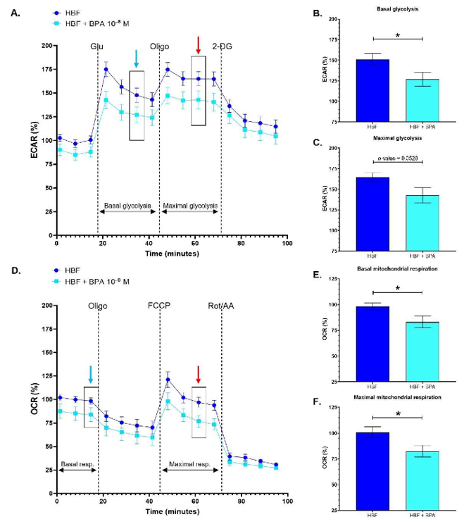
Healthy Human Bladder Fibroblasts Exhibit Decreased Glycolytic and Mitochondrial Metabolism following Chronic Exposure to BPA
BPA is an endocrine disruptor that can bind to cell receptors involved in cancer progression. Here, Pellerin et al. tested the effects of nanomolar concentrations of BPA on the metabolism of bladder fibroblasts and CAFs.
Human bladder fibroblasts (HBFs) were exposed to physiological concentrations of BPA to evaluate the impact of this compound on these cells. In vivo, BPA can reach HBFs through blood vessels nourishing the cells populating the stroma and potentially affects them. HBFs chronically exposed to 10-8 M BPA exhibited a generally decreased energy metabolism compared to untreated HBFs (Fig. 1A, D). HBFs exposed to BPA demonstrated a significantly reduced basal glycolytic metabolism (Fig. 1B), while maximal glycolysis slightly decreased, with a p-value established at 0.0528 (Fig. 1C). HBFs exposed to BPA also exhibited significantly decreased basal and maximal mitochondrial respiration (Fig. 1E, F). Therefore, HBFs chronically exposed to physiological concentrations of BPA are characterized by a reduced glycolytic and mitochondrial oxidative metabolism.
1) The cells are cryopreserved at P2 and marketed as secondary cells to ensure the highest viability and proliferation efficiency. 2) Cells were identified: Fibronectin or Vimentin stained positive by immunofluorescence. 4) Does not contain HIV-1, HBV, HCV, mycoplasma, bacteria, yeast, and fungi.
It is recommended to use SuperCult® Complete Human Fibroblast Medium (cat# CM-1097X) for the culturing of Human Bladder Fibroblasts.
Check all containers for leakage or breakage. Remove the frozen cells from the dry ice packaging and immediately place the cells at a temperature below -130°C, preferably in liquid nitrogen vapor, until ready for use.
Our primary bladder fibroblasts are spindle-shaped and isolated from the urinary bladder of a donor. The cells can be used for the study or development of a potential diagnostic method for the early detection of bladder cancer cells, reconstruction studies, and advancement of cancer research.
Ask a Question
Average Rating: 5.0 | 1 Scientist has reviewed this product
Affordable
We have chosen this product as the most affordable on the market.
11 June 2023
Ease of use
After sales services
Value for money
Write your own review
Description: Creative Bioarray's normal Human Bladder Epithelial Cells - Apex, when grown in LIUro A culture medium, provide an ideal culture model for the study of bladder biology. Cell Features:Human Bladder ...
Description: HBdSMC from Creative Bioarray are isolated from human bladder tissue. HBdSMC are cryopreserved at secondary culture after purification and delivered frozen. Each vial contains >5 x 10^5 cells in 1 ml ...
Description: Human Bladder Microvascular Endothelial Cells from Creative Bioarray are isolated from human bladder tissue. Human Bladder Microvascular Endothelial Cells are grown in T25 tissue culture flasks ...
Description: Human Bladder Epithelial Cells are isolated from normal human bladder tissue.
Description: Creative Bioarray's normal Human Bladder Epithelial Cells - Dome, when grown in LIUro D Complete culture medium, provide an ideal culture model for the study of bladder biology.
Description: HBdSF from Creative Bioarray are isolated from human bladder tissue. HBdSF are cryopreserved at secondary culture (passage one) and are delivered frozen. Each vial contains >5 x 10^5 cells in 1 ml ...