Hamster Kidney Epithelial Cells
Cat.No.: CSC-C9200J
Species: Hamster
Source: Kidney
Cell Type: Epithelial Cell
- Specification
- Background
- Scientific Data
- Q & A
- Customer Review
Hamster Kidney Epithelial Cells are epithelial cells isolated from hamster kidneys. These cells provide an in vitro physiologically relevant model system with which to study kidney function, epithelial biology and kidney-related toxicity. These cells share important structural and functional attributes with renal epithelial cells that make them amenable for studies looking at transport and barrier function, as well as cellular responses to chemical or pharmacological agents.
Hamster Kidney Epithelial Cells possess a classic cobblestone morphology and will proliferate as an adherent monolayer when maintained in standard cell culture conditions. They express epithelial markers as well as several renal-specific transporters responsible for ion exchange, reabsorption of solutes, and xenobiotic transport. These cells can polarize to form epithelial monolayers with functional tight junctions when cultured to confluence. Hamster Kidney Epithelial Cells are frequently utilized in nephrotoxicity assays, as well as in the study of drug transport and sequestration. They are also used to evaluate mechanisms of renal epithelial damage. Moreover, they have been used as part of comparative physiology studies, and to analyze species-specific responses to stimuli as part of hamster disease studies. They have been utilized in viral infection studies.
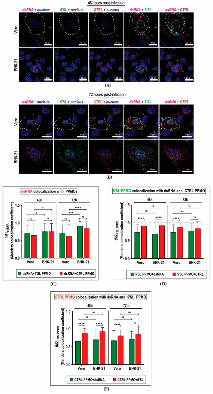
Peptide-Conjugated Phosphorodiamidate Morpholino Oligomers for In Situ Live-Cell Molecular Imaging of Dengue Virus Replication
Non-invasive imaging that preserves spatial and temporal resolution is urgently needed to track pathogens in vivo. Victorio et al. tested whether phosphorodiamidate morpholino antisense oligos coupled to cell-penetrating peptides (PPMOs), previously developed as DENV2 antivirals, could be repurposed as fluorescent in-situ probes for live-cell imaging of DENV2 infection.
They determined whether the 5'SL PPMO specifically tracked DENV2 cellular infection. DENV2-infected Vero and BHK-21 cells were incubated with both PPMOs (10 µM each, 10 min) at 48 h and 72 h post-infection and immunostained for dsRNA, a marker of viral replication intermediates. DENV replication occurs in membrane-bound vesicle packets that appear as intensely fluorescent punctae around nuclei (Fig. 1A, B). To evaluate colocalization with viral replication sites, they calculated the Manders split coefficient (tM1) between dsRNA and either PPMO. The fraction of dsRNA colocalizing with 5'SL PPMO was comparable to that with CTRL PPMO in both cell lines at both time points (Fig. 1C), indicating no selective enrichment of 5'SL PPMO in replication vesicle packets over time. This was expected given that 5'SL and CTRL PPMOs exhibited high mutual colocalization (Fig. 1D, E), exceeding their colocalization with dsRNA. Similar results were observed in ZIKV-infected cells. These findings suggest that most PPMOs either occupied the same vesicles or were trapped in endosomes, rather than entering DENV2 replication compartments.
Ask a Question
Write your own review
Description: Hamster Ovarian Smooth Muscle Cells are isolated from ovarian tissue of pathogen-free laboratory mice.
Description: Hamster Esophageal Epithelial Cells from Creative Bioarray are isolated from esophageal tissue of pathogen-free laboratory mice. Hamster Esophageal Epithelial Cells are grown in a T25 tissue culture ...
Description: Hamster Spleen Epithelial Cells from Creative Bioarray are isolated from spleen tissue of pathogen-free laboratory mice. Hamster Spleen Epithelial Cells are grown in a T25 tissue culture flask ...
Description: Hamster Corneal Epithelial Cells from Creative Bioarray are isolated from corneal tissue of pathogen-free laboratory mice. Hamster Corneal Epithelial Cells are grown in a T25 tissue culture flask ...
Description: Hamster Aortic Smooth Muscle Cells are isolated from aorta of hamster.
Description: Hamster Primary Pancreatic Fibroblasts are isolated from pancreas tissue of hamster.