
Cryopreserved Human Keratinocytes
Cat.No.: CSC-7677W
Species: Human
Source: Epidermis; Skin
Cell Type: Keratinocyte
- Specification
- Background
- Scientific Data
- Q & A
- Customer Review
Cryopreserved human keratinocytes are primary epidermal keratinocytes, typically neonatal or adult, that are expanded, quality-controlled, and stored in liquid nitrogen. After thawing, they recover, attach, and proliferate in serum-free, low-calcium medium, retaining a basal phenotype marked by p63 and cytokeratin 5/14. Upon calcium switch or air-liquid interface culture, they differentiate into stratified epidermis expressing cytokeratin 1/10, involucrin, filaggrin, and loricrin, supporting cornified envelope formation and barrier development.
Their key advantage is readiness for use. Compared with freshly isolated keratinocytes, cryopreserved cells reduce lead time, logistical complexity, and culture failure risk, while enabling flexible scheduling and scalable experiments. Each lot can be qualified for viability, attachment, proliferation, marker expression, and differentiation capacity, improving reproducibility. Compared with immortalized lines such as HaCaT, cryopreserved primary keratinocytes better preserve donor genetic background, native receptor expression, and physiological signaling.
They are widely used in epidermal biology, wound healing, psoriasis, atopic dermatitis, contact dermatitis, photoaging, and skin carcinogenesis, as well as in safety and efficacy testing of drugs, cosmetics, irritants, sensitizers, and wound-care products. They are compatible with 2D monolayers, 3D organotypic skin equivalents, co-culture with fibroblasts or immune cells, and high-content imaging.
The Glycoprotein GPNMB Protects Against Oxidative Stress Through Enhanced PI3K/AKT Signaling in Epidermal Keratinocytes
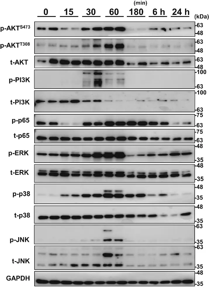
This study aimed to determine the role of glycoprotein nonmetastatic melanoma protein B (GPNMB) in normal and vitiligo epidermis. First, we investigated the intracellular signaling changes after H2O2-induced oxidative stress in cultured normal human epidermal keratinocytes (NHEKs). Then, GPNMB was knocked down to mimic vitiligo keratinocytes (KCs) to investigate the role of GPNMB and its underlying mechanisms in cell susceptibility to H2O2 treatment.
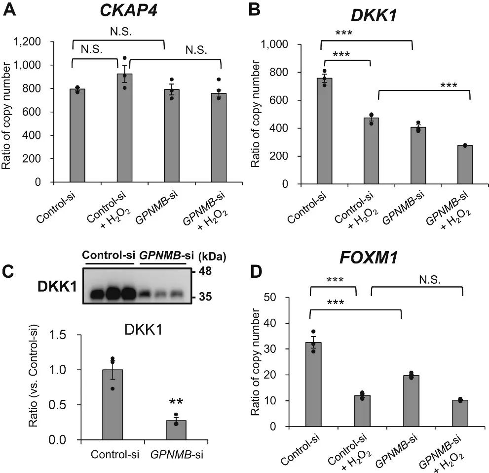
GPNMB knockdown reduced the phosphorylation levels of AKTT308 and AKTS473 after H2O2 treatment, accompanied by reduced Dickkopf-1 (DKK1) mRNA and protein production and decreased FOXM1 mRNA expression. These results suggested that GPNMB protects KCs from H2O2-induced cell death through enhanced PI3K/AKT signaling, and WNT/β-catenin/FOXM1 and DKK1/CKAP4/AKT pathways. Furthermore, a significant increase in thioredoxin-interacting protein (TXNIP) following GPNMB knockdown was observed, indicating the enhanced phosphorylation of JNK and p38 and suppression of WNT/β-catenin signaling. These results suggest that the decreased expression of epidermal GPNMB in vitiligo lesions triggers increased sensitivity to H2O2-induced oxidative stress and decreased WNT/β-catenin signaling, consistent with the pathological features of the vitiligo epidermis.
Ask a Question
Write your own review
Description: Keratinocytes are the primary type of cell found in the epidermis, the outermost layer of the skin. Keratinocytes represent the major cell type of the epidermis, making up about 90% of the cells. ...
Description: Keratinocytes are the primary type of cell found in the epidermis, the outermost layer of the skin. Keratinocytes represent the major cell type of the epidermis, making up about 90% of the cells. ...
Description: Diseased Human Epidermal Keratinocytes (D-HEK) are isolated from human donors that have been diagnosed with diabetes type II disease. Further donor information, including medications, BMI, and ...
Description: Keratinocytes are the primary type of cell found in the epidermis, the outermost layer of the skin. Keratinocytes represent the major cell type of the epidermis, making up about 90% of the cells. ...