
Canine Corneal Epithelial Cells
Cat.No.: CSC-C9113J
Species: Dog
Source: Cornea; Eye
Cell Type: Epithelial Cell
- Specification
- Background
- Scientific Data
- Q & A
- Customer Review
Canine Corneal Epithelial Cells are primary cells isolated from the corneal epithelium of the dog. These cells are the most superficial cell layer of the canine cornea and are comprised of a stratified, non-keratinized epithelial tissue that provides a permeability barrier and protects against mechanical damage, pathogens, and chemical irritants, in addition to facilitating corneal transparency and maintaining homeostasis of the ocular surface. Primary canine corneal epithelial cells maintain many of the morphologic, molecular, and functional features of the parent tissue, offering a physiologically relevant in vitro cell culture model.
The expression of epithelial and cornea-specific markers such as cytokeratins (K3, K12), E-cadherin, and tight junction proteins (ZO-1, claudins) by canine corneal epithelial cells is indicative of their robust barrier function and cellular adhesion. In research settings, these cells are used to investigate the development and differentiation of the cornea, epithelial wound healing and regeneration, inflammatory reactions, and host-pathogen interactions that influence the ocular surface. As dogs naturally present with a high incidence of ocular disorders, such as corneal ulcers, keratoconjunctivitis sicca, and infectious keratitis, canine corneal epithelial cells are a popular cell model to study ophthalmology, perform translational research, and to develop therapeutics in veterinary medicine. Canine corneal epithelial cells are also used as an in vitro testing platform to assess ocular drug permeability, toxicity, and efficacy.
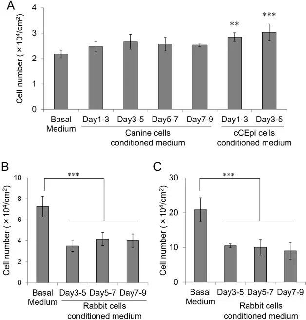
Effect of Conditioned Medium on Corneal Epithelial Cell Growth
Morita et al. hypothesized that canine corneal epithelial cells maintain their proliferative properties through autocrine secretion of growth-promoting factors. They aimed to investigate the maintenance mechanism of the proliferative properties of canine corneal epithelial cells by comparing the autocrine secretion of soluble factors in canine and rabbit corneal epithelial cells.
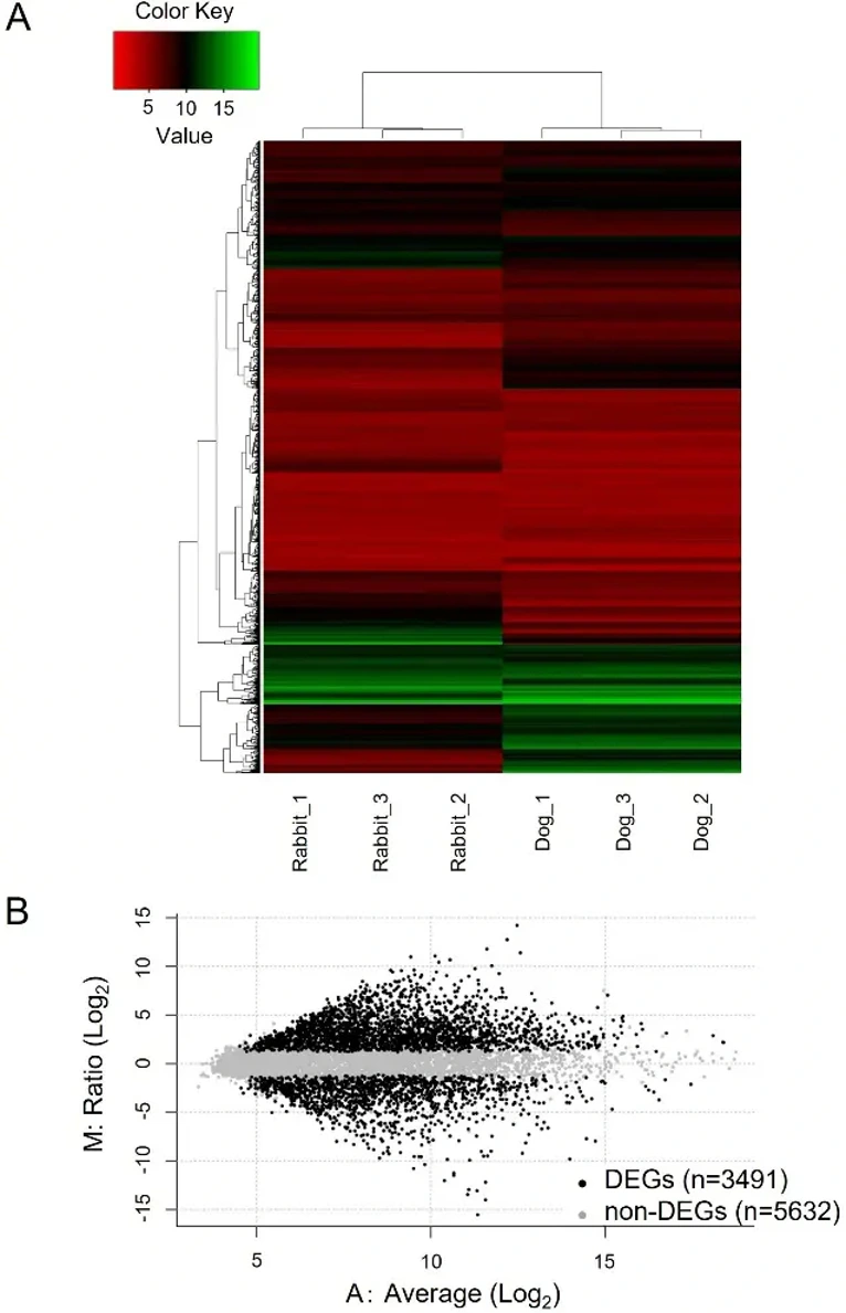
Conditioned media from primary canine corneal epithelial cells slightly increased rabbit cell growth but with no significant differences compared to BM control. In contrast, conditioned media from cCEpi cells significantly promoted rabbit cell growth (Fig. 1A). Conversely, conditioned media from rabbit corneal epithelial cells significantly inhibited the proliferation of both canine and cCEpi cells (Fig. 1B, C). Microarray analysis confirmed experimental validity through hierarchical cluster analysis (Fig. 2A) and identified 3491 DEGs from 9123 annotated genes between canine and rabbit corneal epithelial cells (Fig. 2B). Canine cells had 2156 highly expressed genes, while rabbit cells had 1335. From these, 22 soluble factor genes were selected in canine cells, including four EGFR ligands: NRG1, HBEGF, EREG, and EPGN. In rabbit cells, 12 soluble factor genes were selected with CTGF and TGFB2 being highly expressed.
Ask a Question
Write your own review
Description: Dog Liver Endothelial Cells from Creative Bioarray are isolated from tissue of dog liver. Dog Liver Endothelial Cells are grown in T25 tissue culture flasks pre-coated with gelatin-based coating ...
Description: Canine Astrocytes from Creative Bioarray are isolated from canine brain tissue. The method we use to isolate canine astrocytes were developed based on a combination of established and our proprietary ...
Description: Canine Mammary Microvascular Endothelial Cells from Creative Bioarray are isolated from breast of pathogen-free laboratory Canine. Canine Mammary Microvascular Endothelial Cells are grown in T25 ...
Description: Canine Chondrocytes (CnC) provided by Creative Bioarray are isolated from normal canine articular cartilage tissue. The cells are frozen at passage 1 and each vial contains at least 0.5*10^6 cells. ...
Description: Canine Pancreatic Microvascular Endothelial Cells from Creative Bioarray are isolated from Pancreatic Microvascular of pathogen-free laboratory Canine. Canine Pancreatic Microvascular Endothelial ...
Description: Canine Prostate Microvascular Endothelial Cells from Creative Bioarray are isolated from prostate of pathogen-free laboratory Canine. Canine Prostate Microvascular Endothelial Cells are grown in T25 ...