
CD1 Mouse Lung Microvascular Endothelial Cells
Cat.No.: CSC-C4230X
Species: Mouse
Source: Lung
Cell Type: Endothelial Cell; Microvascular Cell
- Specification
- Scientific Data
- Q & A
- Customer Review
Enhanced Host Neovascularization of Prevascularized Engineered Muscle Following Transplantation into Immunocompetent versus Immunocompromised Mice
Background
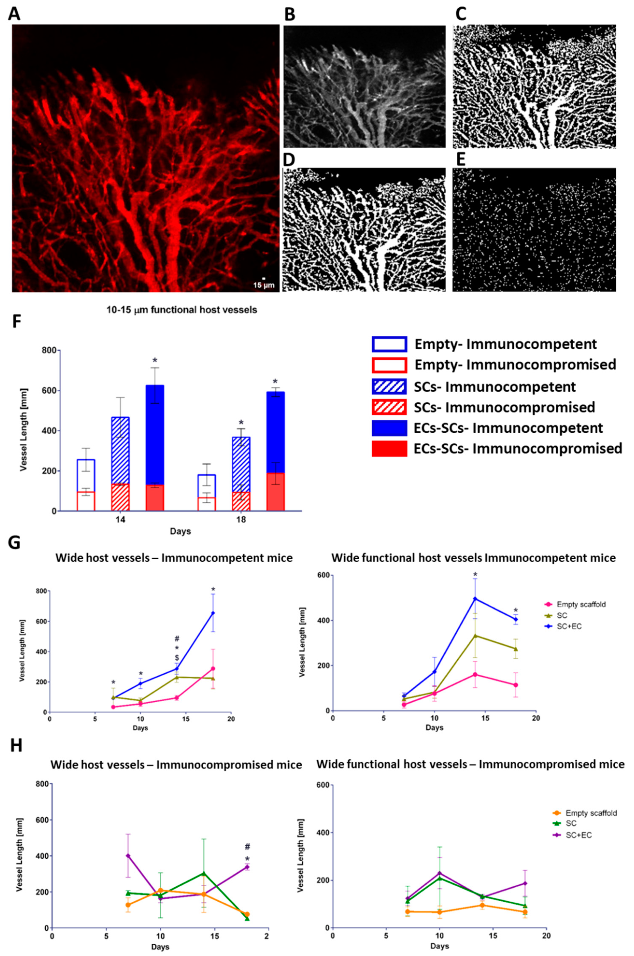
This study engineered a fully murine, prevascularized three-dimensional skeletal muscle construct on PLLA/PLGA scaffolds using endothelial cells and satellite cells, then transplanted it into abdominal wall defects of immunocompetent and immunocompromised mice. Intravital imaging showed that host neovascularization was significantly greater in immunocompetent animals than in immunocompromised controls, especially 14–18 days post-implantation. Prevascularization improved vessel ingrowth, graft integration, and myogenesis in both models. The findings support autologous cell-based tissue engineering over allogeneic approaches and highlight the immune system's beneficial role in vascularizing engineered muscle.
Key Findings
The authors fabricated 3D vascularized murine muscle constructs by co-seeding murine endothelial cells and satellite cells on PLLA/PLGA scaffolds with fibrin gel, and transplanted them into immunocompetent CD1 Flk-1-GFP and immunocompromised athymic nude mice. Intravital imaging through an abdominal window tracked host vessel penetration over 18 days. At days 14 and 18, prevascularized EC+SC grafts in immunocompetent mice had significantly longer functional host vessel networks than grafts in immunocompromised mice, and in both models prevascularized grafts supported more host neovascularization than empty or SC-only scaffolds. EC+SC grafts also showed better integration, greater new myofiber formation, and less extracellular matrix deposition than controls. The study concludes that an intact immune system enhances graft neovascularization and that autologous cell-based prevascularization may be superior to allogeneic transplantation with immunosuppression.
Contribution
Creative Bioarray supplied CD1 mouse lung microvascular endothelial cells used to create the prevascularized engineered muscle constructs. These endothelial cells were co-cultured with satellite cells on PLLA/PLGA scaffolds, enabling vessel-like network formation and in vivo assessment of host neovascularization following transplantation into immunocompetent and immunocompromised mice.
Ask a Question
Write your own review
Description: C57BL/6-GFP Mouse Skeletal Muscle Microvascular Endothelial Cells from Creative Bioarray are isolated from C57BL/6-Tg (CAG-EGFP) 1Osb/J mouse skeletal muscle tissue of pathogen-free laboratory mice. ...
Description: eNOS KO Mouse Stomach Epithelial Cells from Creative Bioarray are isolated from stomach tissue of pathogen-free laboratory mice. eNOS KO Mouse Stomach Epithelial Cells are grown in a T25 tissue ...
Description: eNOS KO Mouse Liver Fibroblasts from Creative Bioarray are isolated from liver tissue of pathogen-free laboratory mice. eNOS KO Mouse Liver Fibroblasts are grown in T75 tissue culture flasks ...
Description: C57BL/6-GFP Mouse Corneal Epithelial Cells from Creative Bioarray are isolated from C57BL/6-GFP-Tg(CAG-EGFP)1Osb/J mouse corneal tissue of pathogen-free laboratory mice. C57BL/6-GFP Mouse Corneal ...
Description: BALB/c Mouse Retinal Microvascular Endothelial Cells from Creative Bioarray are isolated from retinal tissue of pathogen-free laboratory mice. BALB/c Mouse Retinal Microvascular Endothelial Cells are ...