
C57BL/6 Mouse Kidney Glomerular Endothelial Cells
Cat.No.: CSC-C4286X
Species: Mouse
Source: Kidney
Cell Type: Endothelial Cell
- Specification
- Background
- Scientific Data
- Q & A
- Customer Review
C57BL/6 Mouse Kidney Glomerular Endothelial Cells are specialized microvascular endothelial cells derived from renal glomeruli. In vivo, they form the blood-facing component of the glomerular filtration barrier, functioning alongside the glomerular basement membrane and podocytes. Their fenestrated architecture facilitates water and small-solute filtration, while the endothelial surface layer, including the glycocalyx, contributes to restricting plasma protein passage. These properties make glomerular endothelial cells central to investigations of renal filtration and vascular integrity.
A key research advantage is their combination of glomerular tissue origin and an established inbred genetic background. They provide a relevant cellular platform for examining glomerular endothelial mechanisms and facilitate comparisons with genetically modified mouse models.
Appropriately characterized cultures support studies of endothelial permeability, glycocalyx regulation, inflammatory activation, and responses to injury. Mouse glomerular endothelial models have demonstrated increased transendothelial albumin passage following enzymatic disruption of glycocalyx heparan sulfate. Complementary studies in C57BL/6 mice have linked endothelial surface-layer damage to increased glomerular albumin leakage, providing a mechanistic framework for connecting cellular findings with renal vascular outcomes. These applications are particularly valuable for investigating pathways relevant to proteinuria and glomerular barrier dysfunction.
These cells also support investigation of podocyte–endothelial communication, including soluble mediators that influence glycocalyx integrity and barrier function. With validated functional assays, they can help evaluate candidate interventions targeting endothelial injury or restoring barrier properties.
Panax Notoginseng Saponins Attenuate Glomerular Endothelial Glycocalyx Degradation by Inhibiting the HPSE1/EGFR/STAT3 Signaling Pathway
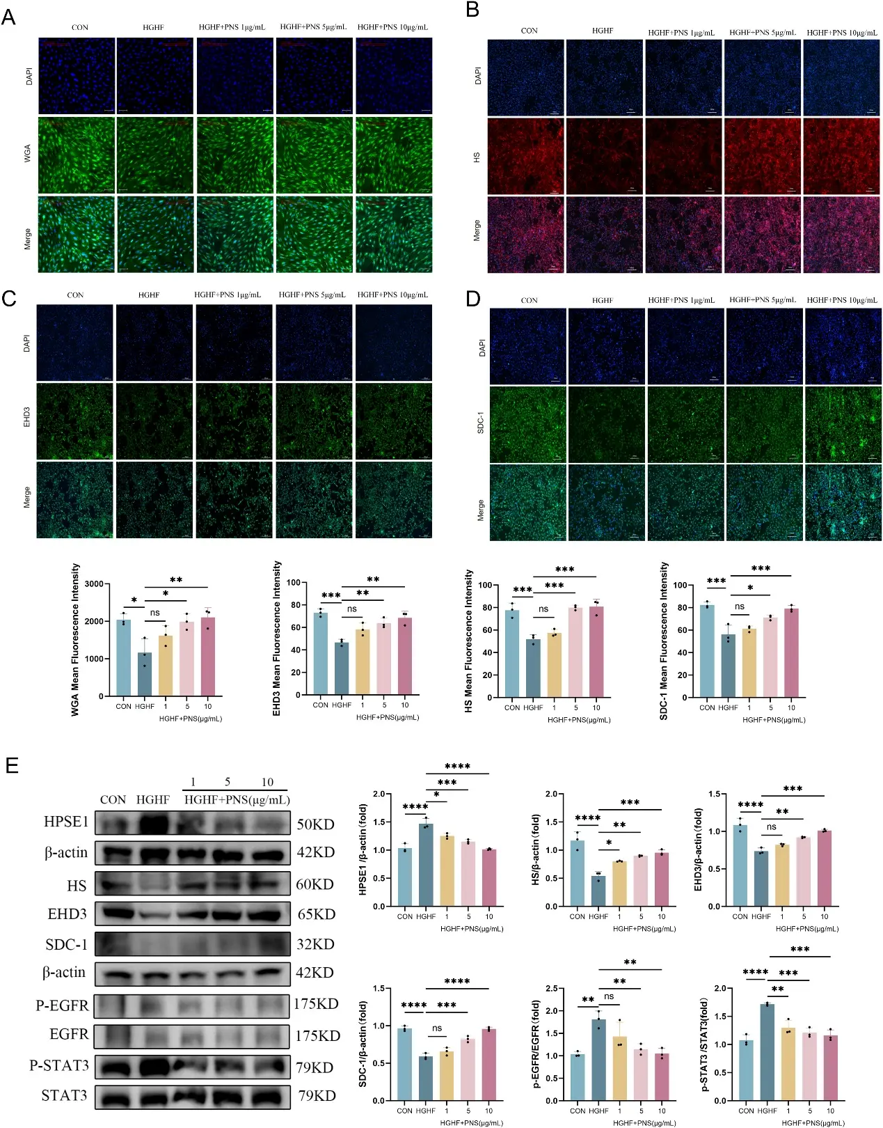
This study investigated whether Panax notoginseng saponins (PNS) protects against glomerular endothelial glycocalyx (eGC) injury in diabetic nephropathy (DN) by regulating the HPSE1/EGFR/STAT3 signaling axis. In vitro cell models of DN were established using mouse renal glomerular endothelial cells (MRGECs) stimulated with high glucose/high palmitic acid (HGHF). WGA staining and the expression of glycocalyx-related components, including HS, SDC-1, and EHD3 were evaluated.
HGHF stimulation reduced WGA fluorescence intensity, indicating loss of glycocalyx staining on the cell surface. Consistently, immunofluorescence and Western blot analyses showed decreased expression of HS, SDC-1, and EHD3 after HGHF exposure. HGHF also increased HPSE1 expression, suggesting enhanced glycocalyx degradation. PNS treatment suppressed HGHF-induced HPSE1 upregulation and restored the levels of HS, SDC-1, and EHD3 to varying degrees (Fig. 1A–E). These results support that PNS helps maintain endothelial glycocalyx integrity under HGHF-induced injury conditions.
Ask a Question
Write your own review
Description: Mouse Pulmonary Microvascular Endothelial Cells are isolated from normal mouse tissue. The cells are characterized by immunofluorescence with antibodies specific to vWF, CD31. T25 flasks is required ...
Description: BALB/c Mouse Spleen Epithelial Cells from Creative Bioarray are isolated from spleen tissue of pathogen-free laboratory mice. BALB/c Mouse Spleen Epithelial Cells are grown in a T25 tissue culture ...
Description: BALB/c Mouse Dermal Endothelial Cells from Creative Bioarray are isolated from skin tissue of pathogen-free laboratory mice. BALB/c Mouse Dermal Endothelial Cells are grown in T25 tissue culture ...
Description: C57BL/6-GFP Mouse Skeletal Muscle Microvascular Endothelial Cells from Creative Bioarray are isolated from C57BL/6-Tg (CAG-EGFP) 1Osb/J mouse skeletal muscle tissue of pathogen-free laboratory mice. ...
Description: eNOS KO Mouse Stomach Epithelial Cells from Creative Bioarray are isolated from stomach tissue of pathogen-free laboratory mice. eNOS KO Mouse Stomach Epithelial Cells are grown in a T25 tissue ...