Mouse Periodontal Ligament Fibroblasts
Cat.No.: CSC-C5412S
Species: Mouse
Source: Periodontal Ligament; Periodontium
Cell Type: Fibroblast
- Specification
- Background
- Scientific Data
- Q & A
- Customer Review
Mouse periodontal ligament fibroblasts from Creative Bioarray are isolated from the mouse tooth tissue. The method we use to isolate mouse periodontal ligament fibroblasts was developed based on a combination of established and our proprietary methods. The mouse periodontal ligament fibroblasts are characterized by immunofluorescence with antibodies specific to vimentin or fibronectin. Each vial contains 0.5x10^6 cells per ml and is delivered frozen.
Mouse Periodontal Ligament Fibroblasts (MPDLFs) are fibroblast-like cells that have been isolated from periodontal ligament (PDL) tissue of mice. The PDL itself is a specialized connective tissue that helps support the tooth from the surrounding alveolar bone and distribute mechanical load during biting and chewing before remodeling tissue components. MPDLFs serve as an in vitro model system for periodontal biology and disease.
Mouse Periodontal Ligament Fibroblasts in culture are spindle-shaped and form adherent monolayers. As expected of fibroblasts, they express vimentin and produce collagen type I, collagen type III and fibronectin. Depending on stimulation conditions, these cells can also express markers associated with osteogenic or cementogenic differentiation, such as alkaline phosphatase (ALP), Runx2, and osteocalcin, reflecting the regenerative potential of periodontal ligament tissue. MPDLFs have been used extensively to study periodontal inflammation and tissue remodeling, orthodontic tooth movement, and alveolar bone regeneration. MPDLFs upregulate proliferation, cytokine secretion, and matrix production when stimulated by mechanical stress, bacterial products (such as lipopolysaccharide), and pro-inflammatory cytokines. Due to these properties they have also been used to study signaling cascades related to inflammation and remodeling such as NF-κB, MAPK and TGF-β.
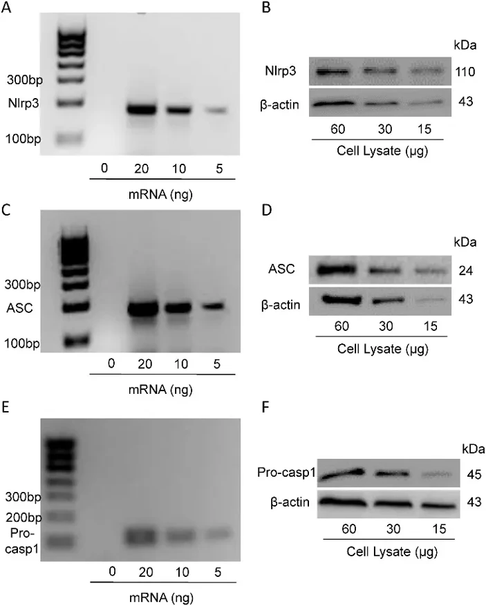
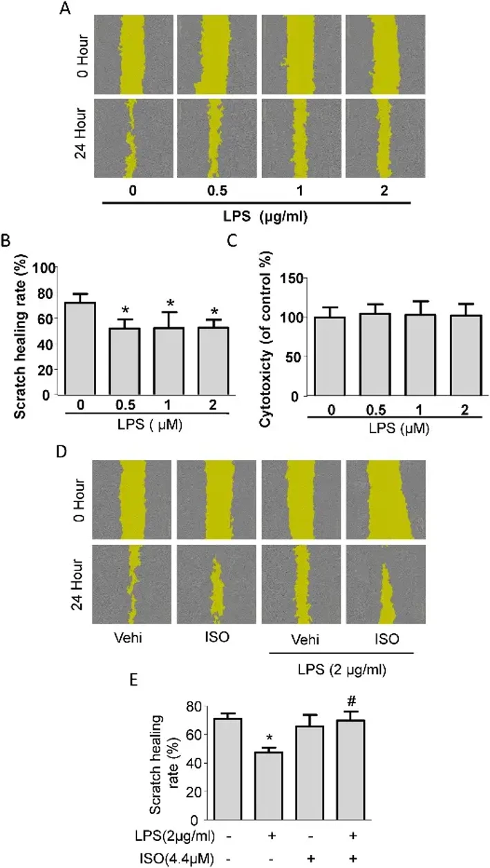
Migration in mPDLFs is Dependent on P.g-LPS Induced Nlrp3 Inflammasome
Inflammasomes initiate inflammatory responses to danger signals, but their role in chronic periodontitis, particularly in periodontal ligament fibroblasts (PDLFs), remains unclear. Lian's team demonstrated abundant Nlrp3 inflammasome component expression in cultured mouse PDLFs (mPDLFs).
RT-PCR and Western blot confirmed mRNA and protein expression of NLRP3, ASC, and Caspase-1 in mPDLFs (Fig. 1A-F). While Porphyromonas gingivalis LPS (P.g-LPS) is known to compromise PDLF functions including migration, its specific regulatory role was unexplored. They found that P.g-LPS markedly decreased mPDLF migration without causing cytotoxicity (Fig. 2A-C). Importantly, the Nlrp3 inflammasome inhibitor ISO recovered P.g-LPS-induced migration dysfunction (Fig. 2D, E), indicating that the Nlrp3 pathway mediates P.g-LPS-induced migration impairment in mPDLFs.
Ask a Question
Write your own review
- You May Also Need

Description: C57BL/6-GFP Mouse Skeletal Muscle Microvascular Endothelial Cells from Creative Bioarray are isolated from C57BL/6-Tg (CAG-EGFP) 1Osb/J mouse skeletal muscle tissue of pathogen-free laboratory mice. ...

Description: eNOS KO Mouse Stomach Epithelial Cells from Creative Bioarray are isolated from stomach tissue of pathogen-free laboratory mice. eNOS KO Mouse Stomach Epithelial Cells are grown in a T25 tissue ...
Description: eNOS KO Mouse Liver Fibroblasts from Creative Bioarray are isolated from liver tissue of pathogen-free laboratory mice. eNOS KO Mouse Liver Fibroblasts are grown in T75 tissue culture flasks ...

Description: C57BL/6-GFP Mouse Corneal Epithelial Cells from Creative Bioarray are isolated from C57BL/6-GFP-Tg(CAG-EGFP)1Osb/J mouse corneal tissue of pathogen-free laboratory mice. C57BL/6-GFP Mouse Corneal ...

Description: BALB/c Mouse Retinal Microvascular Endothelial Cells from Creative Bioarray are isolated from retinal tissue of pathogen-free laboratory mice. BALB/c Mouse Retinal Microvascular Endothelial Cells are ...