Mouse Pancreatic Stellate Cells
Cat.No.: CSC-C5381S
Species: Mouse
Source: Pancreas
Cell Type: Pancreatic Stellate Cell
- Specification
- Background
- Scientific Data
- Q & A
- Customer Review
Mouse pancreatic stellate cells from Creative Bioarray are isolated from the mouse pancreatic tissue. The method we use to isolate mouse pancreatic stellate cells was developed based on a combination of established and our proprietary methods. The mouse pancreatic stellate cells are characterized by immunofluorescence with antibodies specific to desmin or α-SMA. Each vial contains 0.5x10^6 cells per ml and is delivered frozen.
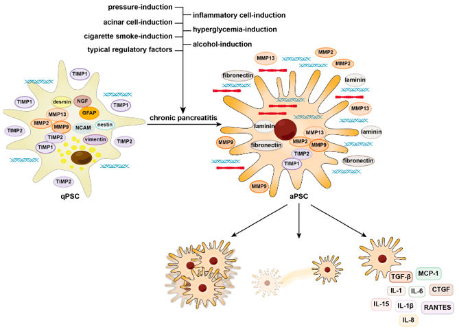
Mouse Pancreatic Stellate Cells (mPSCs) are pancreatic stromal cells originally isolated from mouse pancreas. In the pancreas they are localized to periacinar/periductal areas and have been shown to regulate extracellular matrix (ECM) homeostasis under normal physiological settings. Much like hepatic stellate cells, pancreatic stellate cells are quiescent and contain cytoplasmic vitamin A-containing lipid droplets.
mPSCs become activated upon pancreatic injury or inflammation and can also be activated by exposure to profibrotic mediators like TGF-β or PDGF. When active, they transdifferentiate into myofibroblast-like cells and lose their lipid droplets. Activated mPSCs proliferate and migrate, and express α-smooth muscle actin (α-SMA), vimentin, desmin, and collagen I. Activated cells produce ECM components including collagen, fibronectin, and laminin.
mPSCs have a spindle-shaped fibroblast-like morphology in vitro and proliferate as adherent monolayers. They are frequently used as a tool to study pancreatic fibrosis mechanisms. Their ease of use has also made them valuable in studying chronic pancreatitis and pancreatic ductal adenocarcinoma (PDAC) stromal interactions. mPSCs can be used to study ECM production, cytokine signaling, oxidative stress, and stellate cell-acinar cell, stellate cell-immune cell, and tumor cell-stromal cell communication.
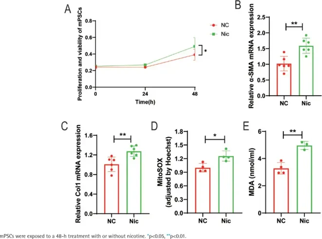
Nicotine Promotes Activation and Induces Mitochondrial Oxidative Stress in mPSCs
Wei's team investigated nicotine effects on pancreatic stellate cell (PSC) activation and pancreatic fibrosis in chronic pancreatitis (CP). Their previous work had demonstrated that treating human PSCs with 1 μM nicotine for 48 hours results in optimal cell activation and mitochondrial oxidative stress. Using these conditions, they exposed mouse pancreatic stellate cells (mPSCs) to nicotine in vitro.
Treatment with nicotine significantly enhanced mPSC activation as shown by increased cell proliferation (Fig. 1A) and upregulated α-SMA mRNA expression (Fig. 1B). Nicotine treatment also activated fibroblasts, as shown by increased mRNA expression of Type I collagen (Col I) (Fig. 1C), pointing towards fibrosis through ECM metabolism disruption. Nicotine enhanced mitochondrial oxidative stress in mPSCs as well. Mitochondrial ROS production was significantly increased compared to controls (Fig. 1D) and malondialdehyde (MDA), one of the products of lipid peroxidation, was also significantly increased (Fig. 1E).
Ask a Question
Write your own review


Description: C57BL/6-GFP Mouse Skeletal Muscle Microvascular Endothelial Cells from Creative Bioarray are isolated from C57BL/6-Tg (CAG-EGFP) 1Osb/J mouse skeletal muscle tissue of pathogen-free laboratory mice. ...

Description: eNOS KO Mouse Stomach Epithelial Cells from Creative Bioarray are isolated from stomach tissue of pathogen-free laboratory mice. eNOS KO Mouse Stomach Epithelial Cells are grown in a T25 tissue ...
Description: eNOS KO Mouse Liver Fibroblasts from Creative Bioarray are isolated from liver tissue of pathogen-free laboratory mice. eNOS KO Mouse Liver Fibroblasts are grown in T75 tissue culture flasks ...
Description: C57BL/6-GFP Mouse Corneal Epithelial Cells from Creative Bioarray are isolated from C57BL/6-GFP-Tg(CAG-EGFP)1Osb/J mouse corneal tissue of pathogen-free laboratory mice. C57BL/6-GFP Mouse Corneal ...

Description: BALB/c Mouse Retinal Microvascular Endothelial Cells from Creative Bioarray are isolated from retinal tissue of pathogen-free laboratory mice. BALB/c Mouse Retinal Microvascular Endothelial Cells are ...