Mouse Oviduct Epithelial Cells
Cat.No.: CSC-C5323S
Species: Mouse
Source: Oviduct
Cell Type: Epithelial Cell
- Specification
- Background
- Scientific Data
- Q & A
- Customer Review
Mouse oviduct epithelial cells from Creative Bioarray are isolated from the mouse oviduct tissue. The method we use to isolate mouse oviduct epithelial cells was developed based on a combination of established and our proprietary methods. The mouse oviduct epithelial cells are characterized by immunofluorescence with antibodies specific to cytokeratin-18 (CK-18). Each vial contains 0.5x10^6 cells per ml and is delivered frozen.
Mouse Oviduct Epithelial Cells (MOECs) are highly specialized cells that line the mammalian oviduct, playing a fundamental role in the early stages of reproduction. These cells provide the essential microenvironment for gamete transport, fertilization, and the first critical divisions of the pre-implantation embryo. Our purified primary MOECs offer a high-fidelity in vitro system that meticulously mirrors the complex physiological signaling of the maternal reproductive tract.
- Ciliated and Secretory Phenotypic Fidelity: MOECs consist of a functional balance of ciliated cells, which facilitate ovum transport, and secretory cells (Peg cells), which produce oviduct-specific glycoproteins (e.g., OVGP1). This dual-cell population is vital for recreating the biochemical "milieu" necessary for successful in vitro fertilization (IVF) and embryo development.
- Superior Model for Reproductive Toxicology: Unlike non-specific cell lines, MOECs retain their sensitivity to hormonal fluctuations (estrogen and progesterone). This renders them an ideal platform for screening the effects of endocrine-disrupting chemicals (EDCs) and pharmacological agents on tubal patency and early embryonic health.
- Indispensable for Pathogenesis Research: These cells serve as a premier model for investigating the origins of pelvic inflammatory disease (PID) and, crucially, the early events in serous ovarian carcinogenesis, as the distal oviduct (fimbria) is increasingly recognized as the primary site for precursor lesions (STIC).
- Enhanced Co-culture Compatibility: Our MOECs are optimized for co-culture systems with gametes or embryos. By secreting embryotrophic factors and neutralizing reactive oxygen species (ROS), they significantly improve embryo quality and blastocyst formation rates compared to synthetic media alone.
Validated for the expression of specific markers such as Cytokeratin 8/18, Acetylated α-tubulin, and PAX8, our Mouse Oviduct Epithelial Cells provide a standardized, "reproduction-ready" platform. They are an essential asset for pharmaceutical R&D, agricultural biotechnology, and academic labs focused on fertility, developmental biology, and gynecological oncology.
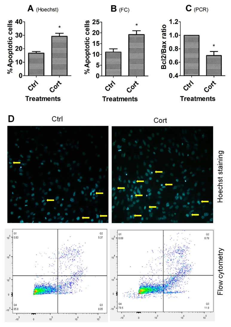
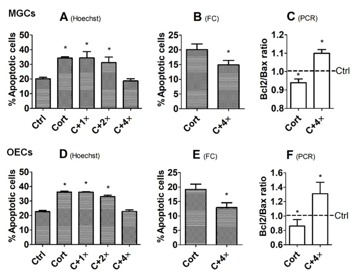
Role of tPA in Corticosterone-Induced Apoptosis of Mouse Mural Granulosa and Oviductal Epithelial Cells
Although studies indicate that female stress-increased secretion of glucocorticoids impairs oocyte competence and embryo development, by inducing apoptosis of ovarian and oviductal cells, respectively, the mechanisms by which glucocorticoids induce apoptosis of ovarian and oviductal cells are largely unclear. Tissue plasminogen activator (tPA) has been involved in apoptosis of different cell types. However, while some studies indicate that tPA is proapoptotic, others demonstrate its antiapoptotic effects. This study has explored the role and action mechanisms of tPA in corticosterone-induced apoptosis of mouse mural granulosa cells (MGCs) and oviductal epithelial cells (OECs).
The results demonstrate that culture with corticosterone significantly increased apoptosis, while decreasing levels of tPA (Plat) mRNA and tPA protein in both MGCs and OECs. Culture with tPA ameliorated corticosterone-induced apoptosis of MGCs and OECs. Furthermore, while tPA protected MGCs from corticosterone-induced apoptosis by interacting with low-density lipoprotein receptor-related protein 1 (LRP1), it protected OECs from the apoptosis by acting on Annexin 2 (ANXA2). In conclusion, tPA is antiapoptotic in both MGCs and OECs, and it protects MGCs and OECs from corticosterone-induced apoptosis by interacting with LRP1 and ANXA2, respectively, suggesting that tPA may use different receptors to inhibit apoptosis in different cell types.
Ask a Question
Write your own review
- You May Also Need

Description: C57BL/6-GFP Mouse Skeletal Muscle Microvascular Endothelial Cells from Creative Bioarray are isolated from C57BL/6-Tg (CAG-EGFP) 1Osb/J mouse skeletal muscle tissue of pathogen-free laboratory mice. ...

Description: eNOS KO Mouse Stomach Epithelial Cells from Creative Bioarray are isolated from stomach tissue of pathogen-free laboratory mice. eNOS KO Mouse Stomach Epithelial Cells are grown in a T25 tissue ...
Description: eNOS KO Mouse Liver Fibroblasts from Creative Bioarray are isolated from liver tissue of pathogen-free laboratory mice. eNOS KO Mouse Liver Fibroblasts are grown in T75 tissue culture flasks ...

Description: C57BL/6-GFP Mouse Corneal Epithelial Cells from Creative Bioarray are isolated from C57BL/6-GFP-Tg(CAG-EGFP)1Osb/J mouse corneal tissue of pathogen-free laboratory mice. C57BL/6-GFP Mouse Corneal ...

Description: BALB/c Mouse Retinal Microvascular Endothelial Cells from Creative Bioarray are isolated from retinal tissue of pathogen-free laboratory mice. BALB/c Mouse Retinal Microvascular Endothelial Cells are ...