
Mouse Cardiomyocytes
Cat.No.: CSC-C5355S
Species: Mouse
Source: Heart
Cell Type: Cardiomyocyte
- Specification
- Background
- Scientific Data
- Q & A
- Customer Review
Mouse cardiomyocytes from Creative Bioarray are isolated from mouse heart tissue. The method we use to isolate mouse cardiomyocytes was developed based on a combination of established and our proprietary methods. The mouse cardiomyocytes from Creative Bioarray are characterized by immunofluorescence with antibodies specific to myosin heavy chain. Each vial contains 0.5x10^6 cells per ml and is delivered frozen.
Mouse cardiomyocytes are cells that make up cardiac muscle tissue. Cardiomyocytes are of particular interest for cardiovascular study as they provide an in vivo look into cardiac contraction, electrophysiology, and disease progression. Mouse cardiomyocytes are often isolated from neonatal or adult mouse heart tissue and can be distinguished by the organized nature of their sarcomeres and dense mitochondrial populations responsible for their rhythmic and involuntary contractions. Neonatal cardiomyocytes are typically preferred for study in the field due to their ability to be more easily cultured and their reduced proliferative ability; however, adult cardiomyocytes are sometimes used as their shape and maturation state more closely represent an in vivo heart. Mouse cardiomyocytes, both neonatal and adult, are used to study calcium handling and signaling, ion channel kinetics, and cardiotoxicity of developing drugs.
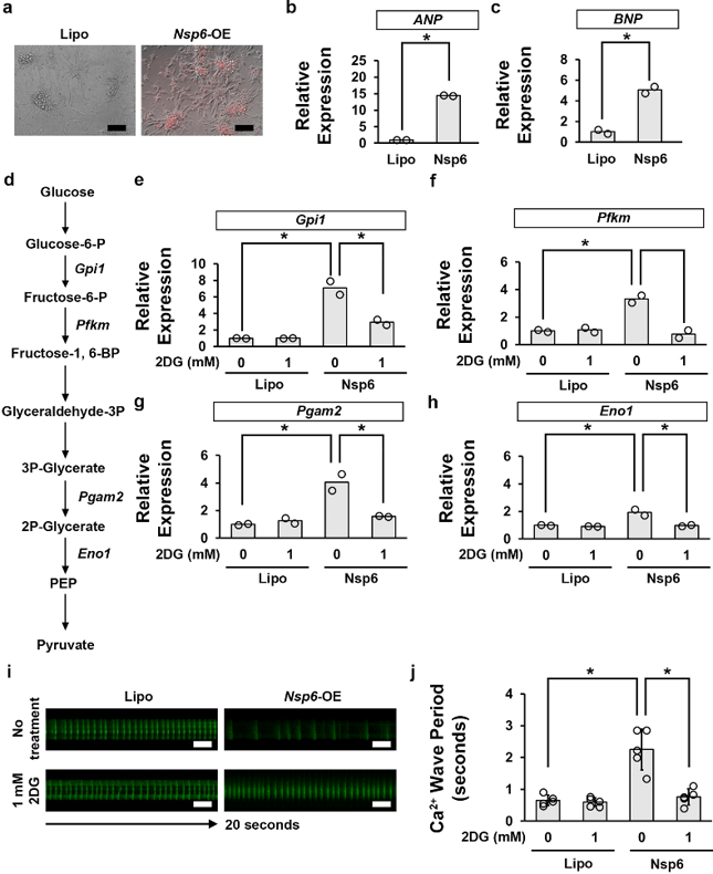
Inhibition of Glycolysis Pathway Activity by 2DG Attenuates SARS-Cov-2 Nsp6-Induced Functional Defect in Mouse Cardiomyocytes
Infection with SARS-CoV-2 leads to COVID-19, a disease characterized by severe respiratory illness and cardiovascular complications. Zhu's team explored the direct impact of SARS-CoV-2 proteins on heart function in Drosophila and mouse models, focusing on a viral protein, Nsp6, that interacts with the host protein MGA/MAX and dysregulates cardiac metabolism.
Nsp6 was found to directly interact with MGA/MAX, which activates glycolysis and disrupts mitochondria, leading to increased ROS. To test whether their observations in flies also hold true in mammals, they isolated heart cells from mouse embryos and added the SARS-CoV-2 Nsp6 gene. They assessed markers of heart cell enlargement (hypertrophy) because dysfunction in cardiac sugar metabolism is known to cause hypertrophy. The Nsp6 gene triggered a rise in ANP and BNP production which indicates cardiac hypertrophy, and they also upregulated genes involved in glycolysis (sugar breakdown). 2DG (a glycolysis inhibitor) treatment rescued all of these changes. Heart cells expressing Nsp6 had prolonged calcium uptake duration, which is important for heart muscle contraction and relaxation. 2DG treatment rescued the calcium uptake duration back to normal levels. Thus, the SARS-CoV-2 protein Nsp6 impairs mouse heart function by altering sugar metabolism and blocking this pathway with 2DG can rescue the heart cell dysfunction.
Ask a Question
Write your own review

Description: C57BL/6-GFP Mouse Skeletal Muscle Microvascular Endothelial Cells from Creative Bioarray are isolated from C57BL/6-Tg (CAG-EGFP) 1Osb/J mouse skeletal muscle tissue of pathogen-free laboratory mice. ...

Description: eNOS KO Mouse Stomach Epithelial Cells from Creative Bioarray are isolated from stomach tissue of pathogen-free laboratory mice. eNOS KO Mouse Stomach Epithelial Cells are grown in a T25 tissue ...

Description: eNOS KO Mouse Liver Fibroblasts from Creative Bioarray are isolated from liver tissue of pathogen-free laboratory mice. eNOS KO Mouse Liver Fibroblasts are grown in T75 tissue culture flasks ...

Description: C57BL/6-GFP Mouse Corneal Epithelial Cells from Creative Bioarray are isolated from C57BL/6-GFP-Tg(CAG-EGFP)1Osb/J mouse corneal tissue of pathogen-free laboratory mice. C57BL/6-GFP Mouse Corneal ...

Description: BALB/c Mouse Retinal Microvascular Endothelial Cells from Creative Bioarray are isolated from retinal tissue of pathogen-free laboratory mice. BALB/c Mouse Retinal Microvascular Endothelial Cells are ...