
Aged Mouse Thymus Epithelial Cells
Cat.No.: CSC-C4669L
Species: Mouse
Source: Thymus
Cell Type: Epithelial Cell
- Specification
- Background
- Scientific Data
- Q & A
- Customer Review
Aged mouse thymus epithelial cells (TECs) are isolated from the involuted thymus of geriatric C57BL/6 mice, faithfully recapitulating the structural and functional hallmarks of age-related thymic degeneration. In contrast to youthful TECs, aged TECs exhibit diminished cortical-medullary organization, reduced AIRE expression, impaired cytokine secretion (e.g., IL-7, TSLP), and increased adipocytic infiltration, mirroring the human immune senescence phenotype.
These cells offer distinct advantages for translational and commercial exploitation. First, they provide a physiologically relevant in vitro model to study thymic involution, failure of central tolerance, and rebound of autoimmune manifestations in aging. Second, aged TECs enable high-throughput screening of rejuvenating agents - including growth hormones, anti-aging compounds, and thymus-regenerative biologics - by quantifying recovery of epithelial integrity, AIRE expression, and T‑cell supportive capacity. Third, they serve as quality-controlled building blocks for senescent thymic organoids and ex vivo drug efficacy platforms, reducing reliance on aged animal cohorts.
Cryopreserved as adherent, phenotypically validated preparations, aged mouse TECs deliver consistent, reproducible performance for aging research, immunotherapeutics, and regenerative medicine. Their commercial value lies in accelerating preclinical validation of interventions targeting immune rejuvenation, thus addressing the growing market for age‑related immune dysfunction and thymic repair.
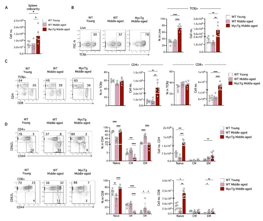
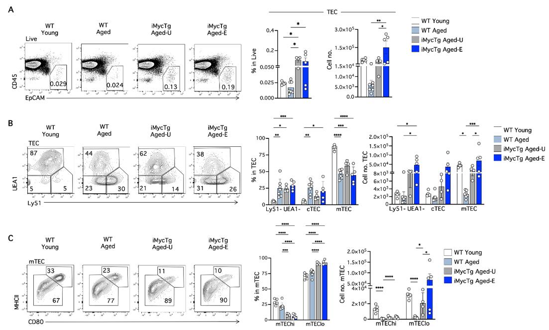
Myc-Mediated TEC Expansion Prevents Age-Associated Alterations to Peripheral T-Cells
Age-related thymic involution leads to diminished output of naïve T-cells. While this process is suggested to increase the risk of disease severity in the elderly following infection, direct evidence is lacking. We developed two mouse models that allow us to experimentally prevent or reverse thymic involution. Constitutive Myc expression in thymic epithelial cells (TEC) of middle-aged mice enhanced thymic function, and increased numbers of peripheral naïve CD4 and CD8 T-cells. Inducible Myc expression reversed age-related thymic involution and partially recovered peripheral naïve T-cell numbers. Importantly, improving thymic function in these settings preserved T-cell-dependent antibody responses and significantly reduced T-cell-associated mortality after infection with Toxoplasma gondii. Improved thymic function also rebalanced age-associated alterations in the Treg pool, and mitigated loss of the transcriptional Th1 signature in aged conventional T-cells. Our findings support the value of TEC-focused thymic regeneration strategies for enhancement of T-cell-mediated immunity in the elderly.
Ask a Question
Write your own review


Description: C57BL/6-GFP Mouse Skeletal Muscle Microvascular Endothelial Cells from Creative Bioarray are isolated from C57BL/6-Tg (CAG-EGFP) 1Osb/J mouse skeletal muscle tissue of pathogen-free laboratory mice. ...

Description: eNOS KO Mouse Stomach Epithelial Cells from Creative Bioarray are isolated from stomach tissue of pathogen-free laboratory mice. eNOS KO Mouse Stomach Epithelial Cells are grown in a T25 tissue ...

Description: eNOS KO Mouse Liver Fibroblasts from Creative Bioarray are isolated from liver tissue of pathogen-free laboratory mice. eNOS KO Mouse Liver Fibroblasts are grown in T75 tissue culture flasks ...
Description: C57BL/6-GFP Mouse Corneal Epithelial Cells from Creative Bioarray are isolated from C57BL/6-GFP-Tg(CAG-EGFP)1Osb/J mouse corneal tissue of pathogen-free laboratory mice. C57BL/6-GFP Mouse Corneal ...

Description: BALB/c Mouse Retinal Microvascular Endothelial Cells from Creative Bioarray are isolated from retinal tissue of pathogen-free laboratory mice. BALB/c Mouse Retinal Microvascular Endothelial Cells are ...