
CD1 Mouse Pancreatic Islets
Cat.No.: CSC-C9292J
Species: Mouse
Source: Pancreatic Islet; Pancreas
- Specification
- Background
- Scientific Data
- Q & A
- Customer Review
CD1 Mouse Pancreatic Islets are isolated islets of Langerhans from CD1 mice. Islets are primary cell clusters cultured from mouse pancreases. They are used as an in vitro model of pancreatic biology that is physiologically relevant to glucose homeostasis and diabetes. These pancreatic clusters consist of multiple hormone-secreting cells types including insulin producing β cells, glucagon-producing α cells, somatostatin-secreting δ cells, as well as pancreatic polypeptide (PP) cells.
CD1 mouse pancreatic islets express markers characteristic of pancreatic hormone producing cells such as insulin, glucagon, and Pdx1. They maintain a 3-dimensional morphology when in culture. CD1 Mouse pancreatic islets are responsive to glucose as well as other secretagogues. They can be used for various assays to determine insulin release as well as understand β-cell function and islet physiology. When maintained in culture, CD1 Mouse pancreatic islets can be viable for several days. Islets are used to model diabetes and understand glucose homeostasis. These islets can be used in applications such as islet transplantation, metabolic regulation, and β-cell toxicity. Because they are primary cells from mice, CD1 Mouse pancreatic islets have utility as a preclinical model. The CD1 mouse is an outbred strain, so pancreatic islets derived from this strain will have genetic diversity.
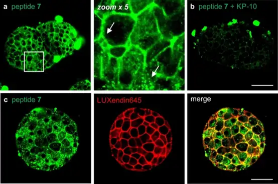
Fluorescence Imaging of GPR54 Distribution in Intact Mouse Pancreatic Islets
The kisspeptin receptor GPR54 regulates reproduction, metabolism and cancer, yet lacks reliable fluorescent tools for live-cell and tissue imaging. Mendive-Tapia et al. designed an acid-resistant BODIPY amino acid (Trp-BODIPY PLUS) to create fluorogenic kisspeptin probes that enable wash-free visualization of GPR54.
GPR54 is widely expressed in hypothalamic and peripheral tissues including liver and pancreas, where kisspeptin regulates insulin secretion from β cells and plays a key role in glucose metabolism. Pancreatic islets contain primarily insulin-secreting β cells alongside glucagon-secreting α cells and somatostatin-producing δ cells. Since β cell loss or dysfunction contributes to both type 1 and type 2 diabetes, and kisspeptin/GPR54 expression is downregulated in diabetic models, this system represents a potential therapeutic target for restoring β cell function. To image GPR54 distribution in pancreatic islets, they incubated intact islets from wild-type CD1 mice with fluorogenic agonistic peptide 7 (10 μM) for 1 h. Confocal microscopy revealed bright membrane labeling and intracellular puncta, indicating GPR54 expression and ligand-mediated internalization (Fig. 1a). Specificity was confirmed by competition with unlabeled KP-10, which substantially reduced membrane fluorescence, demonstrating comparable binding affinity of both peptides for GPR54 (Fig. 1b).
Ask a Question
Write your own review
- You May Also Need

Description: C57BL/6-GFP Mouse Skeletal Muscle Microvascular Endothelial Cells from Creative Bioarray are isolated from C57BL/6-Tg (CAG-EGFP) 1Osb/J mouse skeletal muscle tissue of pathogen-free laboratory mice. ...

Description: eNOS KO Mouse Stomach Epithelial Cells from Creative Bioarray are isolated from stomach tissue of pathogen-free laboratory mice. eNOS KO Mouse Stomach Epithelial Cells are grown in a T25 tissue ...
Description: eNOS KO Mouse Liver Fibroblasts from Creative Bioarray are isolated from liver tissue of pathogen-free laboratory mice. eNOS KO Mouse Liver Fibroblasts are grown in T75 tissue culture flasks ...

Description: C57BL/6-GFP Mouse Corneal Epithelial Cells from Creative Bioarray are isolated from C57BL/6-GFP-Tg(CAG-EGFP)1Osb/J mouse corneal tissue of pathogen-free laboratory mice. C57BL/6-GFP Mouse Corneal ...

Description: BALB/c Mouse Retinal Microvascular Endothelial Cells from Creative Bioarray are isolated from retinal tissue of pathogen-free laboratory mice. BALB/c Mouse Retinal Microvascular Endothelial Cells are ...