
C57BL/6 Mouse Vein Endothelial Cells
Cat.No.: CSC-C4234X
Species: Mouse
Source: Vein
Cell Type: Endothelial Cell
- Specification
- Background
- Scientific Data
- Q & A
- Customer Review
These C57BL/6 Mouse Vein Endothelial Cells (MVECs) are primary endothelial cells, sourced from the venous system of C57BL/6 mice, a common choice in biomedical research. Molecularly, MVECs can be confirmed by canonical endothelial markers such as CD31 (PECAM-1), von Willebrand Factor (vWF) and VE-cadherin. Functionally, MVECs have been shown to take up acetylated low-density lipoprotein (Ac-LDL) and form capillary-like structures in Matrigel-based tube formation assays.
As compared to arterial endothelial cells, mouse vein endothelial cells are ideal for studying leukocyte trafficking as the venous system represents where trans-endothelial migration occurs during inflammation. Additionally, they can be used as an in vitro model to consistently quantify cytokine (TNF-α, IL-1β, etc.) induced expression of adhesion molecules such as ICAM-1 and VCAM-1. These cells can also be used as a consistent in vitro model to study vascular permeability, endothelial metabolism, and murine-specific mechanisms of angiogenesis.
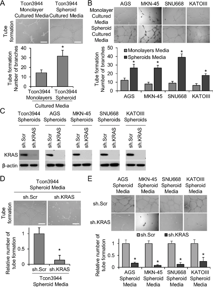
Conditioned Media from Gastric Cancer Spheroid Cells Promotes Angiogenesis
The previous work demonstrated that KRAS activation in gastric cancer cells induces epithelial-to-mesenchymal transition (EMT) and generates cancer stem-like cells (CSCs). Here, Yoon et al. investigated how KRAS activation in gastric CSCs promotes tumor angiogenesis and metastasis. Gastric CSCs were found to secrete pro-angiogenic factors such as VEGF-A, and KRAS inhibition markedly reduced their secretion.
They utilized a genetically engineered mouse model of gastric adenocarcinoma (GA) driven by Trp53 loss, Cdh1 loss, and oncogenic KrasG12D in gastric parietal cells (Tcon mice). These mice developed intestinal and diffuse-type GAs with 100% penetrance and metastases to lymph nodes, lung, and liver. Tumor-derived organoids and cell lines (Tcon3077 and Tcon3944) were generated from two Tcon tumors. As spheroid culture enriches for CSCs, they compared conditioned media from Tcon3944 monolayer versus spheroid cells in tube formation assays with mouse vein endothelial cells (MVEC). Spheroid-conditioned media induced 2.2-fold greater tube formation than monolayer-conditioned media (Fig. 1A), with similar results for Tcon3077 cells. Human GA cell lines (AGS, MKN-45, SNU668, KATOIII) grown as spheroids similarly showed 1.9-4.3-fold increased HUVEC tube formation compared to monolayers (Fig. 1B). To determine if KRAS drives angiogenic factor secretion, KRAS was knocked down using shRNA (efficiency confirmed by Western blot; Fig. 1C). KRAS knockdown in Tcon3944 spheroid cells reduced MVEC tube formation by 86.3% (Fig. 1D), with comparable results in Tcon3077 cells and human GA spheroid cells (75-91% reduction; Fig. 1E). These data indicate that KRAS activation in GA spheroid cells promotes pro-angiogenic factor secretion.
Ask a Question
Write your own review
Description: C57BL/6-GFP Mouse Skeletal Muscle Microvascular Endothelial Cells from Creative Bioarray are isolated from C57BL/6-Tg (CAG-EGFP) 1Osb/J mouse skeletal muscle tissue of pathogen-free laboratory mice. ...

Description: eNOS KO Mouse Stomach Epithelial Cells from Creative Bioarray are isolated from stomach tissue of pathogen-free laboratory mice. eNOS KO Mouse Stomach Epithelial Cells are grown in a T25 tissue ...

Description: eNOS KO Mouse Liver Fibroblasts from Creative Bioarray are isolated from liver tissue of pathogen-free laboratory mice. eNOS KO Mouse Liver Fibroblasts are grown in T75 tissue culture flasks ...

Description: C57BL/6-GFP Mouse Corneal Epithelial Cells from Creative Bioarray are isolated from C57BL/6-GFP-Tg(CAG-EGFP)1Osb/J mouse corneal tissue of pathogen-free laboratory mice. C57BL/6-GFP Mouse Corneal ...

Description: BALB/c Mouse Retinal Microvascular Endothelial Cells from Creative Bioarray are isolated from retinal tissue of pathogen-free laboratory mice. BALB/c Mouse Retinal Microvascular Endothelial Cells are ...