
C57BL/6 Mouse Retinal Microvascular Endothelial Cells
Cat.No.: CSC-C4238X
Species: Mouse
Source: Retina; Eye
Cell Type: Endothelial Cell; Microvascular Cell
- Specification
- Background
- Scientific Data
- Q & A
- Customer Review
C57BL/6 mouse retinal microvascular endothelial cells (RMECs) are primary endothelial cells isolated from the retinal vasculature of C57BL/6 inbred mice. They retain canonical endothelial markers—CD31/PECAM-1, VE-cadherin, VEGFR2, and CD34—and key blood-retinal barrier (BRB) proteins, including claudin-5, occludin, and ZO-1. Functionally, they exhibit VEGF-responsive proliferation, migration, and tube formation, as well as barrier properties measurable by TEER and permeability assays. As non-immortalized cells, they better preserve native retinal endothelial signaling, junctional integrity, and inflammatory responses than immortalized lines.
Their defined C57BL/6 genetic background provides excellent compatibility with syngeneic C57BL/6 models, including oxygen-induced retinopathy, streptozotocin-induced diabetic retinopathy, retinal neovascularization, and transgenic/knockout strains. This strain matching reduces allogeneic variability and supports mechanistic studies of retinal angiogenesis, BRB breakdown, leukocyte adhesion, oxidative stress, and inflammation. Applications include target validation, drug screening, and toxicity testing for anti-VEGF agents, corticosteroids, and metabolic or inflammatory modulators.
The Mechanism of TNF-α Combined With High Glucose in Regulating Calnexin Aggravates Endothelial Cell Injury of Diabetic Retinopathy
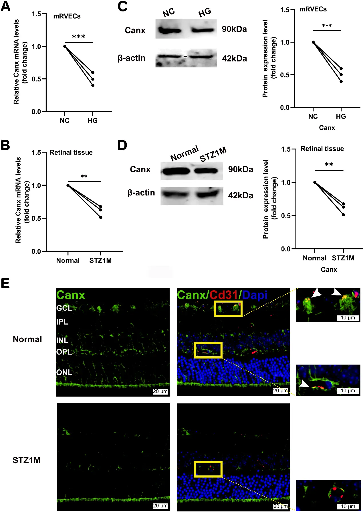
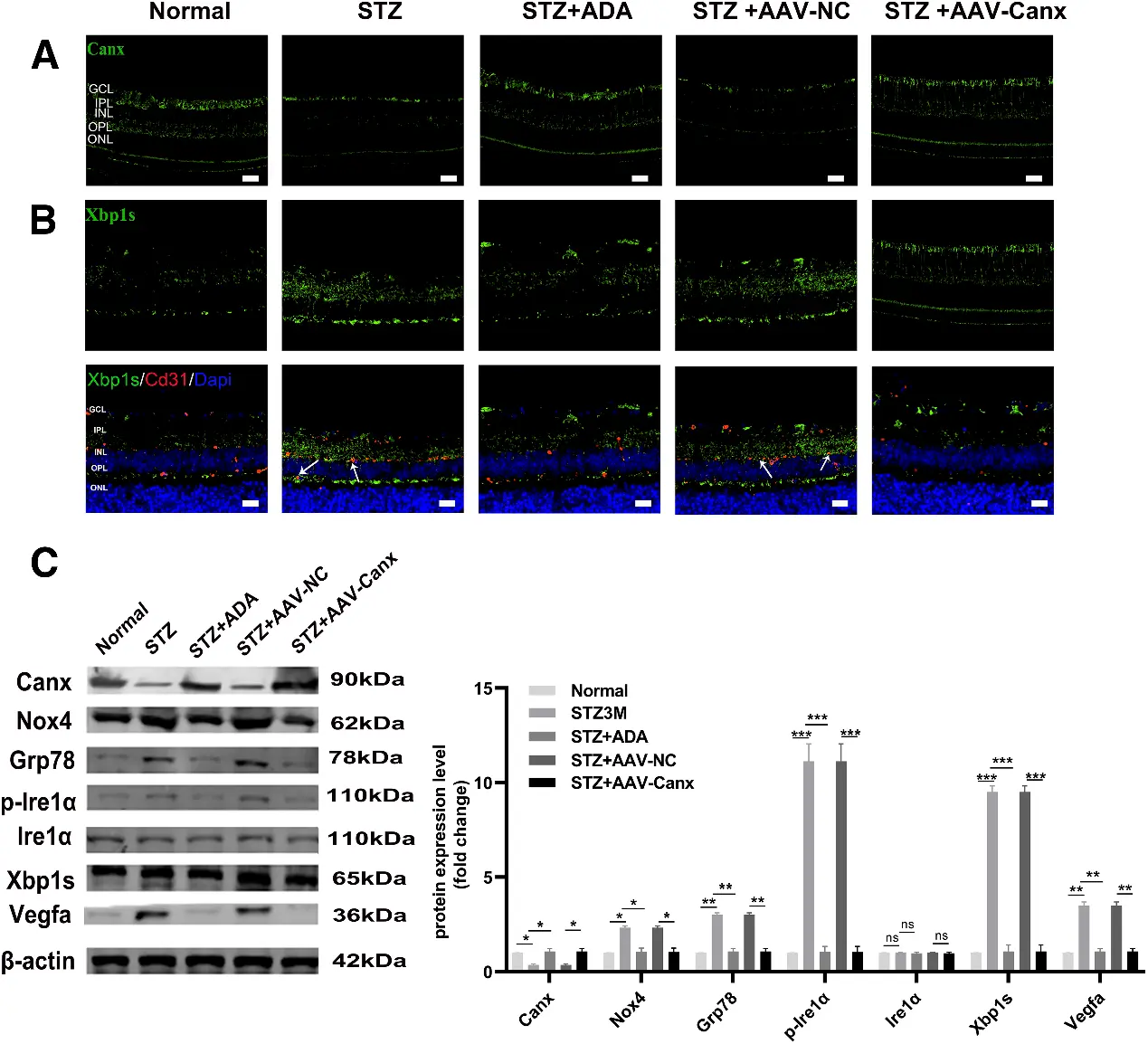
Diabetic retinopathy (DR) is a major cause of irreversible vision loss driven primarily by retinal vascular damage, yet its mechanisms remain incompletely understood. Here, we identify calnexin (Canx) as a critical suppressor of pathological angiogenesis in DR. We demonstrate that hyperglycemia synergizes with TNF-α to downregulate Canx in mouse retinal microvascular endothelial cells. This loss of Canx activates Nox4, leading to hyperactivation of the Ire1α/Xbp1s branch of the unfolded protein response. Consequently, endoplasmic reticulum stress is amplified, pathological Vegfa transcription is upregulated, and the inner blood-retinal barrier is disrupted.
In streptozotocin-induced diabetic mouse models, Canx deficiency exacerbated endothelial dysfunction and retinal vascular pathology. Conversely, both adalimumab (ADA) treatment and adeno-associated virus–mediated Canx overexpression in vivo suppressed the Nox4/Ire1α/Xbp1s/Vegfa cascade, significantly reduced vascular leakage and acellular capillary formation, attenuated retinal thinning, and normalized endothelial cell functions (proliferation, migration, tube formation). Collectively, our findings establish Canx as a key upstream regulator of Vegfa-mediated vascular injury in DR.
Ask a Question
Write your own review
Description: Mouse Pulmonary Microvascular Endothelial Cells are isolated from normal mouse tissue. The cells are characterized by immunofluorescence with antibodies specific to vWF, CD31. T25 flasks is required ...
Description: BALB/c Mouse Spleen Epithelial Cells from Creative Bioarray are isolated from spleen tissue of pathogen-free laboratory mice. BALB/c Mouse Spleen Epithelial Cells are grown in a T25 tissue culture ...
Description: BALB/c Mouse Dermal Endothelial Cells from Creative Bioarray are isolated from skin tissue of pathogen-free laboratory mice. BALB/c Mouse Dermal Endothelial Cells are grown in T25 tissue culture ...
Description: C57BL/6-GFP Mouse Skeletal Muscle Microvascular Endothelial Cells from Creative Bioarray are isolated from C57BL/6-Tg (CAG-EGFP) 1Osb/J mouse skeletal muscle tissue of pathogen-free laboratory mice. ...
Description: eNOS KO Mouse Stomach Epithelial Cells from Creative Bioarray are isolated from stomach tissue of pathogen-free laboratory mice. eNOS KO Mouse Stomach Epithelial Cells are grown in a T25 tissue ...