C57BL/6 Mouse Primary Kidney Epithelial Cells
Cat.No.: CSC-C4265X
Species: Mouse
Source: Kidney
Cell Type: Epithelial Cell
- Specification
- Background
- Scientific Data
- Q & A
- Customer Review
Mouse Primary Kidney Epithelial Cells can be used in assays of cell to cell adhesion and migration. Standard biochemical procedures performed with epithelial cell cultures include RT-PCR, Western blotting, immunoprecipitation, immunofluorescent staining or immunofluorescent flow cytometry or generating cell derivatives for desired research applications.
C57BL/6 Mouse Primary Kidney Epithelial Cells are non-immortalized primary cells isolated from the renal tissue of pathogen-free C57BL/6 inbred mice, a widely used genetic background in biomedical research. Derived from the renal cortex and medulla, these cells primarily consist of proximal and distal tubular epithelial cells, retaining the morphological and functional characteristics of native kidney epithelial cells in vivo. They exhibit a typical polygonal, cobblestone-like epithelial morphology when adherent, and express specific phenotypic markers including pan-cytokeratin (Pan-CK), cytokeratin 18 (CK18), E-cadherin, and aquaporin 1 (AQP1), which confirm their epithelial identity and renal origin.
In vitro, these primary cells have a limited lifespan, typically passable for 2-4 generations and surviving for 1-2 weeks, requiring specialized kidney epithelial cell medium supplemented with growth factors (e.g., EGF, insulin) and culture flasks pre-coated with collagen or fibronectin to promote adherence and proliferation. As a physiologically relevant model, they are widely applied in studies of renal epithelial function, kidney injury and repair, renal inflammation, drug-induced nephrotoxicity, and the pathogenesis of renal diseases such as acute kidney injury and chronic kidney disease.
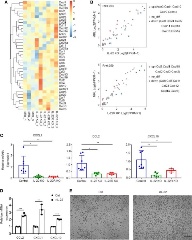
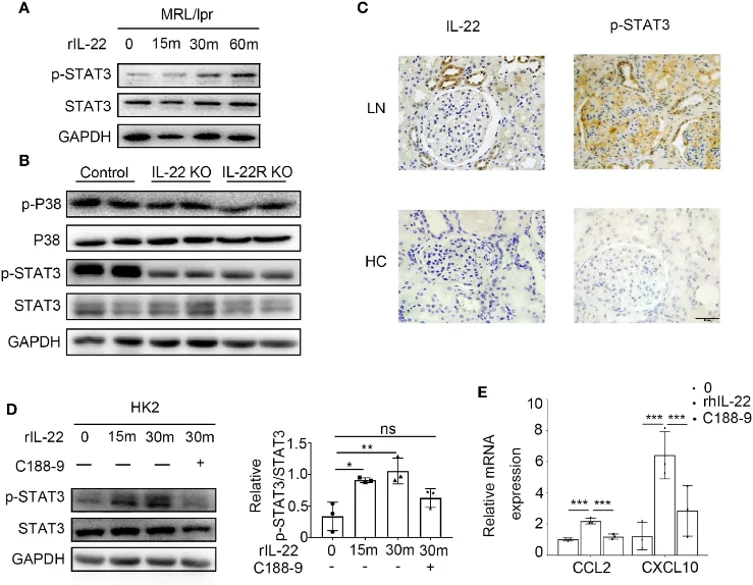
IL-22 Promoted Kidney Epithelial Cells to Express CCL2 and CXCL10 by Activating the STAT3 Pathway
Lupus nephritis (LN) causes severe renal damage in systemic lupus erythematosus patients, with IL-22 implicated in pathogenesis. Hu's team used IL-22 and IL-22R knockout MRL/lpr mice, RNA-sequencing, immunoblotting, and in vitro assays with recombinant IL-22 on primary kidney epithelial cells and HK2 cells to elucidate mechanisms.
IL-22 from type 3 innate lymphoid cells activated STAT3 signaling in kidney epithelial cells, upregulating CCL2 and CXCL10 to recruit macrophages, thereby aggravating LN. Immunoblotting showed significantly increased phosphorylated STAT3 in primary mouse kidney epithelial cells treated with rIL-22 (Fig. 2A). In vivo, STAT3 phosphorylation was markedly higher in control mice at 24 weeks compared to IL-22 KO or IL-22R KO mice, with no difference in p38 (Fig. 2B). Immunochemistry confirmed elevated phosphorylated STAT3 and IL-22 in LN patient kidneys versus healthy controls (Fig. 1C). Similar results were observed in HK2 cells. Moreover, STAT3 inhibitor C188-9 pretreatment significantly reduced rIL-22-induced CCL2 and CXCL10 mRNA expression in HK2 cells (Fig. 1D, E).
Ask a Question
Write your own review

Description: C57BL/6-GFP Mouse Skeletal Muscle Microvascular Endothelial Cells from Creative Bioarray are isolated from C57BL/6-Tg (CAG-EGFP) 1Osb/J mouse skeletal muscle tissue of pathogen-free laboratory mice. ...

Description: eNOS KO Mouse Stomach Epithelial Cells from Creative Bioarray are isolated from stomach tissue of pathogen-free laboratory mice. eNOS KO Mouse Stomach Epithelial Cells are grown in a T25 tissue ...
Description: eNOS KO Mouse Liver Fibroblasts from Creative Bioarray are isolated from liver tissue of pathogen-free laboratory mice. eNOS KO Mouse Liver Fibroblasts are grown in T75 tissue culture flasks ...
Description: C57BL/6-GFP Mouse Corneal Epithelial Cells from Creative Bioarray are isolated from C57BL/6-GFP-Tg(CAG-EGFP)1Osb/J mouse corneal tissue of pathogen-free laboratory mice. C57BL/6-GFP Mouse Corneal ...

Description: BALB/c Mouse Retinal Microvascular Endothelial Cells from Creative Bioarray are isolated from retinal tissue of pathogen-free laboratory mice. BALB/c Mouse Retinal Microvascular Endothelial Cells are ...