
C57BL/6 Mouse Peritoneal Macrophages (Fresh Cells)
Cat.No.: CSC-C4279X
Species: Mouse
Source: Peritoneal Cavity
Cell Type: Macrophage
- Specification
- Background
- Scientific Data
- Q & A
- Customer Review
Cells can be expanded on a multiwell culture plate ready for experiments under the cell culture conditions specified by Creative Bioarray. Repeated freezing and thawing of cells is not recommended.
Mouse Peritoneal Macrophages are tested for expression of markers using antibodies, CD11b by flow cytometry.
Standard biochemical procedures performed with cell cultures include RT-PCR, Western blotting, immunoprecipitation, immunofluorescent staining, flow cytometry or generating cell derivatives for desired research applications.
C57BL/6 mouse peritoneal macrophages are primary innate immune cells isolated from the peritoneal cavity of the widely used C57BL/6 inbred strain. These cells serve as sentinels of host defense, exhibiting potent phagocytic, antigen-presenting, and immunomodulatory functions. They are capable of classical (M1) or alternative (M2) polarization in response to microbial stimuli, cytokines, or tissue injury, thereby mirroring key aspects of inflammation, resolution, and tissue repair. Primary peritoneal macrophages retain physiological receptor expression (TLR2/4/7, FcγR, complement receptors), authentic cytokine profiles (IL-1β, TNF-α, IL-6, IL-10, TGF-β), and intact signaling cascades, ensuring higher predictive power for in vivo outcomes.
Cryopreserved preparations of C57BL/6 peritoneal macrophages are characterized by high viability (>90%), defined surface markers (F4/80+, CD11b+, CD68+, and inducible MHC-II), and functional purity. They enable high-throughput screening of immunomodulators, anti-inflammatory compounds, and nanomedicines, as well as phagocytosis assays, pathogen-host interaction studies, and macrophage polarization profiling. Batch-to-batch reproducibility reduces experimental variability and animal usage, accelerating preclinical drug discovery and immune cell therapy validation. These cells represent a premium, ready-to-use tool for pharmaceutical, biotechnology, and contract research organizations (CROs) targeting chronic inflammatory and immune-mediated diseases.
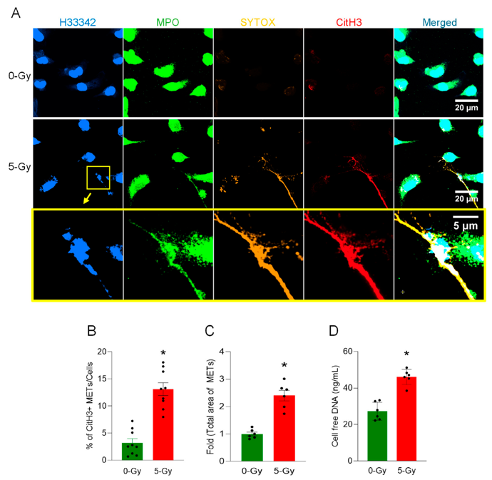
Ionizing Radiation Induces Extracellular Trap Release from Macrophages
Macrophages are key innate immune cells in the host defense against pathogens. Ionizing radiation can impair macrophage functions such as phagocytosis and activate them, potentially exacerbating tissue injury. Macrophage extracellular traps (METs) are formed upon stimulation of macrophages with PAMPs (pathogen-associated molecular pattern) or DAMPs (damage-associated molecular pattern). We hypothesized that macrophages exposed to ionizing radiation can release extracellular traps.
Peritoneal macrophages were collected from C57BL/6 mice and subjected to 5 Gy radiation. We performed assays to detect METs, including the immunofluorescence of citrullination of histone H3 (CitH3) and cell-free DNA measurement in cell culture medium as well as cell death. The results demonstrated MET formation in irradiated macrophages compared to the control (Fig. 1A). The proportion of CitH3 over the total number of nuclei in the microscopic fields was significantly elevated upon the exposure of 5 Gy radiation from 3.2 to 13.1% (Fig. 1B). The measurement of the absolute number of CitH3-positive DNA segments also showed a 3.5-fold increase compared to that of non-irradiated control cells. Total MET area with 5 Gy radiation was increased 2.4-fold compared to the control cell culture (Fig. 1C). We also measured the concentration of cell-free DNA, which is free-floating DNA in the cell culture media. The concentration of cell-free DNA was increased to 170% by the irradiation (Fig. 1D). These data show that radiation increases CitH3 and MPO, which are frequently used as MET markers.
Ask a Question
Write your own review


Description: C57BL/6-GFP Mouse Skeletal Muscle Microvascular Endothelial Cells from Creative Bioarray are isolated from C57BL/6-Tg (CAG-EGFP) 1Osb/J mouse skeletal muscle tissue of pathogen-free laboratory mice. ...

Description: eNOS KO Mouse Stomach Epithelial Cells from Creative Bioarray are isolated from stomach tissue of pathogen-free laboratory mice. eNOS KO Mouse Stomach Epithelial Cells are grown in a T25 tissue ...

Description: eNOS KO Mouse Liver Fibroblasts from Creative Bioarray are isolated from liver tissue of pathogen-free laboratory mice. eNOS KO Mouse Liver Fibroblasts are grown in T75 tissue culture flasks ...
Description: C57BL/6-GFP Mouse Corneal Epithelial Cells from Creative Bioarray are isolated from C57BL/6-GFP-Tg(CAG-EGFP)1Osb/J mouse corneal tissue of pathogen-free laboratory mice. C57BL/6-GFP Mouse Corneal ...

Description: BALB/c Mouse Retinal Microvascular Endothelial Cells from Creative Bioarray are isolated from retinal tissue of pathogen-free laboratory mice. BALB/c Mouse Retinal Microvascular Endothelial Cells are ...