
C57BL/6 Mouse Lymphatic Endothelial Cells
Cat.No.: CSC-C1870
Species: Mouse
Source: Lymph Node
Cell Type: Endothelial Cell
- Specification
- Background
- Scientific Data
- Q & A
- Customer Review
C57BL/6 mouse lymphatic endothelial cells (LECs) are primary lymphatic endothelial cells isolated from lymphatic-rich tissues of the C57BL/6 inbred strain, including lymph nodes, dermis, lung, or mesentery. They retain canonical lymphatic markers—LYVE-1, podoplanin (PDPN), PROX1, and VEGFR-3 (FLT4)—and key functions such as VEGF-C/VEGFR-3–dependent proliferation, migration, tube formation in three-dimensional matrices, and responsiveness to inflammatory cytokines and shear stress. As non-immortalized primary cells, they better preserve native signaling, transcriptional profiles, and strain-specific responses than immortalized LEC lines.
Compared with xenogeneic systems, C57BL/6 LECs provide a defined genetic background and excellent compatibility with syngeneic C57BL/6 mouse models, immune cells, and tumor lines. This reduces allogeneic variability and supports mechanistic studies of lymphangiogenesis, immune cell trafficking, antigen transport, and lipid homeostasis. They are widely used in cancer metastasis, lymphedema, chronic inflammation, autoimmune disease, transplant rejection, and drug discovery. Their defined background also facilitates comparison with widely used knockout, transgenic, and reporter models.
Decorin Evokes a Pro-Lysosomal Pathway in Lymphatic Endothelial Cells
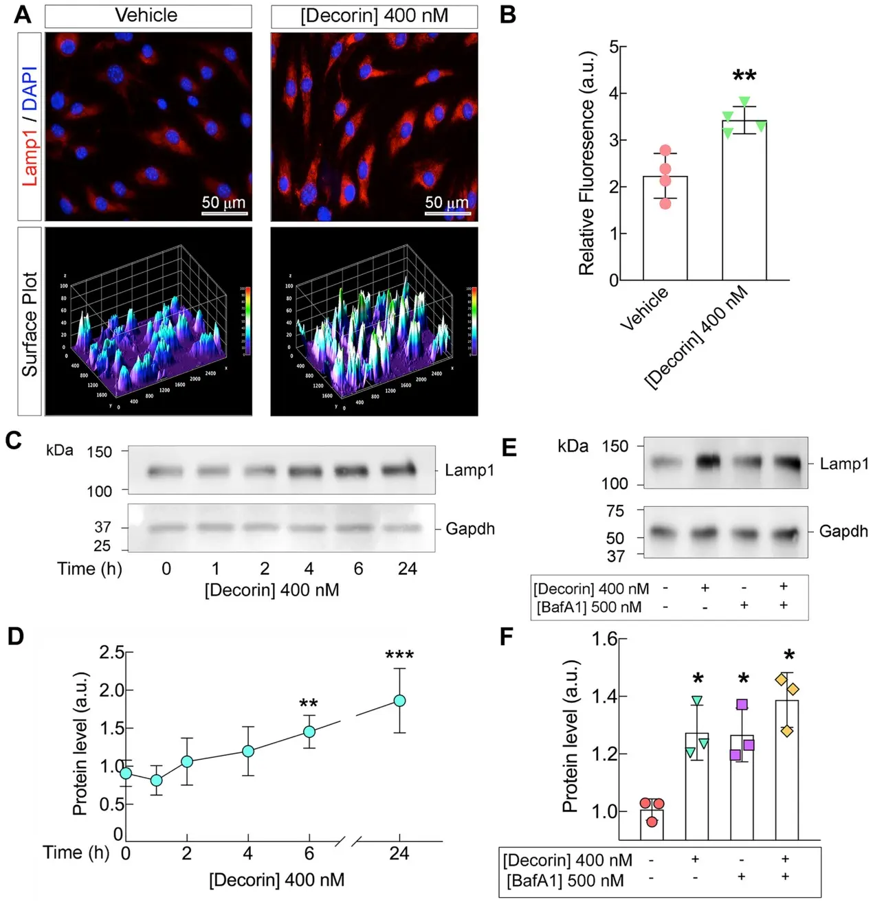
The lymphatic system is critical to the body's immune and circulatory system, and lymphangiogenesis, the development of new lymphatic vessels from pre-existing ones, is a significant process capitalized upon by cancer during tumorigenesis. Decorin is a small leucine-rich proteoglycan which we have previously shown to be anti-tumorigenic and a suppressor of lymphangiogenesis. We have also shown that decorin exercises its anticancer properties through its ability to evoke autophagy. Through a comprehensive and unbiased proteomic analysis, we explored the implications of decorin exposure on protein expression within mouse lymphatic endothelial cells. We discovered that decorin enriches several protein pathways, notably proteasomal degradation and lysosomal pathways. Several proteins within these pathways such as lysosome associated membrane protein 1 (Lamp1) and Neural precursor cell expressed developmentally downregulated protein 8 (Nedd8) were differentially regulated following decorin treatment. These proteins and their functional pathways should be considered therapeutic targets and emerge as candidates for further exploration within the context of decorin and cancer suppression.
C57BL/6 Mouse Lymphatic Endothelial Cells from Creative Bioarray are tested for expression of markers using antibody, VE-cadherin, AF1002, CD31/PECAM-1 or LYVE-1 by immunofluorescence staining or FACS.
Cells can be expanded for 3-6 passages at a split ratio of 1:2 under the cell culture conditions specified by Creative Bioarray.
Ask a Question
Write your own review
Description: Mouse Pulmonary Microvascular Endothelial Cells are isolated from normal mouse tissue. The cells are characterized by immunofluorescence with antibodies specific to vWF, CD31. T25 flasks is required ...
Description: BALB/c Mouse Spleen Epithelial Cells from Creative Bioarray are isolated from spleen tissue of pathogen-free laboratory mice. BALB/c Mouse Spleen Epithelial Cells are grown in a T25 tissue culture ...
Description: BALB/c Mouse Dermal Endothelial Cells from Creative Bioarray are isolated from skin tissue of pathogen-free laboratory mice. BALB/c Mouse Dermal Endothelial Cells are grown in T25 tissue culture ...
Description: C57BL/6-GFP Mouse Skeletal Muscle Microvascular Endothelial Cells from Creative Bioarray are isolated from C57BL/6-Tg (CAG-EGFP) 1Osb/J mouse skeletal muscle tissue of pathogen-free laboratory mice. ...
Description: eNOS KO Mouse Stomach Epithelial Cells from Creative Bioarray are isolated from stomach tissue of pathogen-free laboratory mice. eNOS KO Mouse Stomach Epithelial Cells are grown in a T25 tissue ...