
C57BL/6-GFP Mouse Colonic Epithelial Cells
Cat.No.: CSC-C9091J
Species: Mouse
Source: Colon; Intestine
Cell Type: Epithelial Cell
- Specification
- Background
- Scientific Data
- Q & A
- Customer Review
C57BL/6-GFP Mouse Colonic Epithelial Cells are primary epithelial cells derived from the colon of C57BL/6 mice that constitutively express green fluorescent protein (GFP). The endogenous expression of GFP enables direct visualization and tracking of these cells in various in vitro assays and in vivo-related studies. With the stable and bright GFP signal, cell morphology, viability, proliferation, and migration can be monitored in real time without additional labeling or staining, offering a convenient and practical advantage for imaging-based assays, co-culture systems, and other applications.
The cells display GFP expression while keeping essential features of colonic epithelium through both polarized epithelial shape and expression of specific markers including E-cadherin alongside cytokeratins and tight junction proteins. When cultured under suitable conditions, these cells can form cohesive epithelial layers with barrier-related properties, making them useful for studies on intestinal permeability, epithelial renewal, and host-microenvironment interactions. Applications of C57BL/6-GFP mouse colonic epithelial cells span a wide range of research areas, including intestinal inflammation, colorectal disease and injury, epithelial regeneration and renewal, and gut-immune crosstalk. The C57BL/6 genetic background of these cells ensures compatibility with a broad range of transgenic and disease models, while GFP expression facilitates cell tracing in co-culture studies, organoid integration, and transplantation or injury-repair experiments.
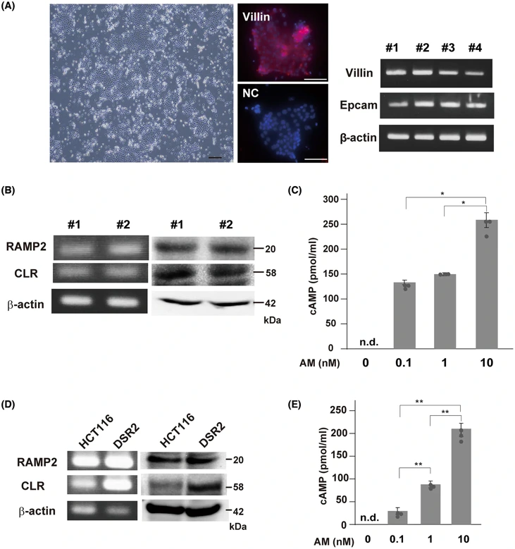
Adrenomedullin Alleviates Mucosal Injury in Experimental Colitis and Increases Claudin-4 Expression in the Colonic Epithelium
Adrenomedullin (AM) is an anti-inflammatory peptide in the intestine mucosa, but its detailed mechanism and effects on intestinal epithelial cells are still unclear. Kawaguchi et al. investigated AM's effects on the expression of junctional molecules in primary-cultured murine intestinal epithelial cells.
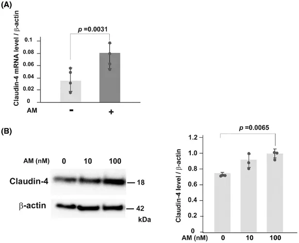
First, the authors cultured murine colonic epithelial cells and confirmed epithelial identity by detecting Villin, Vil1, and Epcam (Fig. 1A). Murine colonic epithelial cells expressed AM receptor molecules RAMP2 and CLR, but not RAMP3 (Fig. 1B). Furthermore, AM treatment increased intracellular cAMP level in a dose-dependent manner (Fig. 1C). Similarly, human colon epithelial cell line HCT116 expressed RAMP2 and CLR, but not RAMP3 and showed cAMP accumulation after AM treatment (Fig. 1D and Fig. 1E). They next investigated whether AM affects the expression of cell adhesion molecules in murine colonic epithelial cells. AM treatment upregulated several cell adhesion-related genes, such as Cldn4 that encodes claudin-4. Upregulation of claudin-4 mRNA and protein levels were confirmed by RT-PCR (Fig. 2A) and immunoblotting (Fig. 2B).
Ask a Question
Write your own review
Description: C57BL/6-GFP Mouse Skeletal Muscle Microvascular Endothelial Cells from Creative Bioarray are isolated from C57BL/6-Tg (CAG-EGFP) 1Osb/J mouse skeletal muscle tissue of pathogen-free laboratory mice. ...

Description: eNOS KO Mouse Stomach Epithelial Cells from Creative Bioarray are isolated from stomach tissue of pathogen-free laboratory mice. eNOS KO Mouse Stomach Epithelial Cells are grown in a T25 tissue ...

Description: eNOS KO Mouse Liver Fibroblasts from Creative Bioarray are isolated from liver tissue of pathogen-free laboratory mice. eNOS KO Mouse Liver Fibroblasts are grown in T75 tissue culture flasks ...
Description: C57BL/6-GFP Mouse Corneal Epithelial Cells from Creative Bioarray are isolated from C57BL/6-GFP-Tg(CAG-EGFP)1Osb/J mouse corneal tissue of pathogen-free laboratory mice. C57BL/6-GFP Mouse Corneal ...

Description: BALB/c Mouse Retinal Microvascular Endothelial Cells from Creative Bioarray are isolated from retinal tissue of pathogen-free laboratory mice. BALB/c Mouse Retinal Microvascular Endothelial Cells are ...