
BALB/c Mouse Proximal Tubular Epithelial Cells
Cat.No.: CSC-C9075J
Species: Mouse
Source: Kidney
Cell Type: Epithelial Cell
- Specification
- Background
- Scientific Data
- Q & A
- Customer Review
BALB/c mouse proximal tubular epithelial cells (mPTECs) are primary cells isolated from the renal cortex of the inbred BALB/c mouse strain. These cells exhibit the characteristic cobblestone-like epithelial morphology and form confluent monolayers with typical dome formation, indicating the presence of tight junctions and an intact transcellular transport process. Immunofluorescent characterization confirms high purity, with more than 95% of cells positive for the epithelial marker cytokeratin 18 (CK18) and over 90% expressing proximal tubule-specific markers including villin, aquaporin-1 (AQP1), and sodium-glucose cotransporter 2 (SGLT2). Additional markers such as E-cadherin and ZO-1 further validate the epithelial phenotype.
The primary advantage of BALB/c mPTECs lies in their retention of in vivo-like differentiated functions-a feature lost in immortalized cell lines. These cells preserve critical renal functions including ion transport, filtration-related activities, and cellular responses to nephrotoxic stimuli. Under optimized culture conditions on collagen-coated membranes, the cells form structurally polarized epithelia with numerous microvilli, basolateral invaginations, and apical tight junctions. Electrophysiological measurements reveal a low transepithelial resistance and high short-circuit current carried by Na+, closely recapitulating the "leaky epithelium" phenotype characteristic of proximal tubules in vivo.
AMPK Protects Proximal Tubular Epithelial Cells from Lysosomal Dysfunction and Dedifferentiation Induced by Lipotoxicity
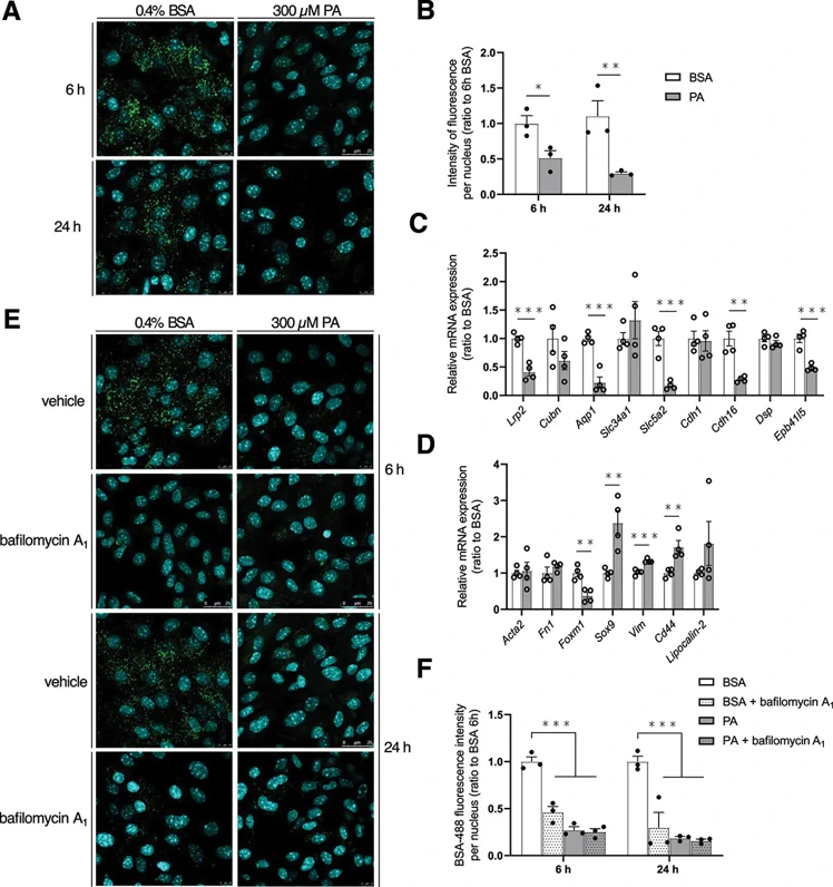
AMP-activated protein kinase (AMPK) was highlighted as a key mediator in the development and progression of obesity-induced chronic kidney disease (CKD). The enzyme is abundantly expressed in kidneys, but its renal activity is decreased in response to metabolic stresses. To further investigate how this pathway is affected upon lipid challenge, primary Mus musculus (mouse) proximal tubular epithelial cells (MmPTECs) were exposed to palmitate (PA), widely used to mimic lipotoxicity in vitro. As soon as 6 h after palmitate exposure, cells displayed impaired lysosomal acidification subsequently leading to autophagosome accumulation and activation of lysosomal biogenesis. We also showed the inability of lysosomal quality control to restore acidic pH which finally drove PTECs dedifferentiation. When PA-induced AMPK activity decline was prevented by AMPK activators, lysosomal acidification and the differentiation profile of PTECs were preserved. Our work provided key insights on the importance of lysosomes in PTECs homeostasis and lipotoxicity and demonstrated the potential of AMPK in protecting the organelle from lipid stress.
Ask a Question
Write your own review

Description: C57BL/6-GFP Mouse Skeletal Muscle Microvascular Endothelial Cells from Creative Bioarray are isolated from C57BL/6-Tg (CAG-EGFP) 1Osb/J mouse skeletal muscle tissue of pathogen-free laboratory mice. ...
Description: eNOS KO Mouse Stomach Epithelial Cells from Creative Bioarray are isolated from stomach tissue of pathogen-free laboratory mice. eNOS KO Mouse Stomach Epithelial Cells are grown in a T25 tissue ...
Description: eNOS KO Mouse Liver Fibroblasts from Creative Bioarray are isolated from liver tissue of pathogen-free laboratory mice. eNOS KO Mouse Liver Fibroblasts are grown in T75 tissue culture flasks ...

Description: C57BL/6-GFP Mouse Corneal Epithelial Cells from Creative Bioarray are isolated from C57BL/6-GFP-Tg(CAG-EGFP)1Osb/J mouse corneal tissue of pathogen-free laboratory mice. C57BL/6-GFP Mouse Corneal ...
Description: BALB/c Mouse Retinal Microvascular Endothelial Cells from Creative Bioarray are isolated from retinal tissue of pathogen-free laboratory mice. BALB/c Mouse Retinal Microvascular Endothelial Cells are ...