

BALB/c Mouse Primary Embryonic Fibroblasts
Cat.No.: CSC-C4325X
Species: Mouse
Source: Embryo
Cell Type: Fibroblast
- Specification
- Background
- Scientific Data
- Q & A
- Customer Review
Mouse Primary Embryonic Fibroblasts are negative for bacteria, yeast, fungi, and mycoplasma. Cells are tested for expression of marker using the antibody of anti-FSP1/S100A4 by immunofluorescence staining. Cells can be expanded for 2-4 passages at a split ratio of 1:2 under the cell culture conditions specified by Creative Bioarray. Repeated freezing and thawing of cells is not recommended.
Standard biochemical procedures performed with cell cultures include the assay of cell to cell interaction, RT-PCR, Western blotting, immunoprecipitation, immunofluorescent staining, flow cytometry or generating cell derivatives for desired research applications.
Primary BALB/c mouse embryonic fibroblasts (BALB/c MEFs), isolated from embryos at day 13.5–14.5 of gestation, are a genetically defined, non-immortalized stromal cell population that serves as an indispensable tool in stem cell biology, cancer research, and immunology.
A principal strength of BALB/c MEFs lies in their H-2d haplotype, which is shared with widely employed BALB/c-derived tumor lines such as 4T1 mammary carcinoma and CT26 colon carcinoma. This enables co-culture and co-injection studies in fully immunocompetent hosts without confounding allogeneic rejection, providing a faithful platform for investigating tumor–fibroblast crosstalk, extracellular matrix remodeling, and immune exclusion within a matched genetic background. As primary cells, they retain a normal diploid karyotype, exhibit stringent contact inhibition, and undergo replicative senescence after a limited number of passages, making them an authentic normal counterpart for oncogenic transformation and senescence bypass experiments. Mitotically inactivated BALB/c MEFs, prepared by mitomycin C treatment or gamma irradiation, serve as robust feeder layers that efficiently support the self-renewal and pluripotency of mouse embryonic stem cells and induced pluripotent stem cells, secreting essential factors such as LIF and extracellular matrix components. Additionally, the inherent Th2-biased immune milieu of the BALB/c strain renders these fibroblasts particularly valuable for studying fibrotic and allergic cytokine responses.
Stress Response of CDK8/19 in Mouse Embryonic Fibroblasts
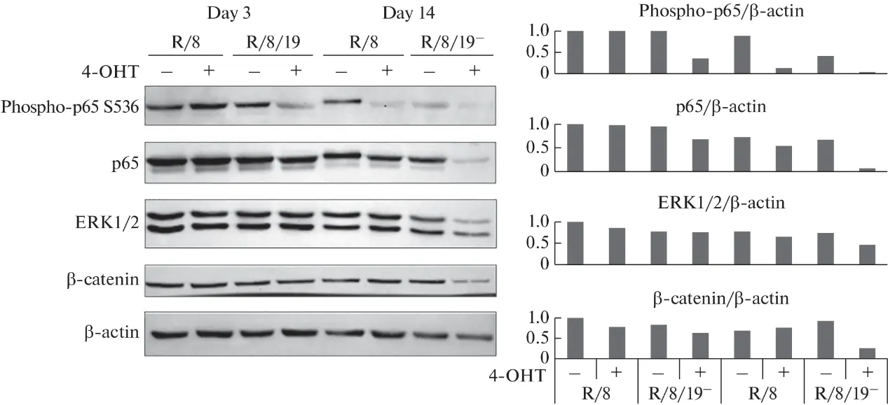
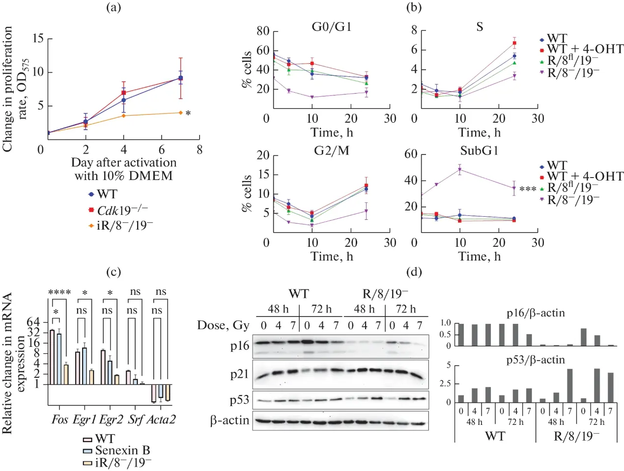
CDK8/19 are primarily necessary for transcriptional reprogramming, that is, a transition from one state with stable expression to another, or short-time induction of expression. The effects of a gene knockout or inhibition of CDK8/19 are therefore expected to occur mostly during activation of transcriptional programs or transcriptional responses to stimuli and stress factors. Varlamova, E. A., et al. chose mouse embryonic fibroblasts (MEFs) to perform an initial search for such effects. MEFs provide an ex vivo model that is most often used for the purpose and are cells that are capable not only of differentiating into various tissues, but also to produce many responses that are not observed in postnatal cells.
The results indicate that CDK8/19 activity is necessary for long-term MEF proliferation and survival and that a deletion of the two kinase genes in vitro distorts the expression regulation of genes involved in many crucial processes, such as metabolism, cell adhesion, and the anti-inflammatory response, and is capable of affecting the potential differentiation direction (adipogenesis and osteogenesis). A Cdk8/Cdk19 knockout causes substantial changes in several signaling pathways (TNF, Wnt, and ERK1/2); reduces the levels of active NF-κB and a phosphorylated form of the retinoblastoma protein (pRB) in long-term cultures; and decreases the cell response to stimuli, for example, when cells exit quiescence or respond to DNA damage.
Ask a Question
Write your own review
Description: C57BL/6-GFP Mouse Skeletal Muscle Microvascular Endothelial Cells from Creative Bioarray are isolated from C57BL/6-Tg (CAG-EGFP) 1Osb/J mouse skeletal muscle tissue of pathogen-free laboratory mice. ...
Description: eNOS KO Mouse Stomach Epithelial Cells from Creative Bioarray are isolated from stomach tissue of pathogen-free laboratory mice. eNOS KO Mouse Stomach Epithelial Cells are grown in a T25 tissue ...
Description: eNOS KO Mouse Liver Fibroblasts from Creative Bioarray are isolated from liver tissue of pathogen-free laboratory mice. eNOS KO Mouse Liver Fibroblasts are grown in T75 tissue culture flasks ...
Description: C57BL/6-GFP Mouse Corneal Epithelial Cells from Creative Bioarray are isolated from C57BL/6-GFP-Tg(CAG-EGFP)1Osb/J mouse corneal tissue of pathogen-free laboratory mice. C57BL/6-GFP Mouse Corneal ...
Description: BALB/c Mouse Retinal Microvascular Endothelial Cells from Creative Bioarray are isolated from retinal tissue of pathogen-free laboratory mice. BALB/c Mouse Retinal Microvascular Endothelial Cells are ...