
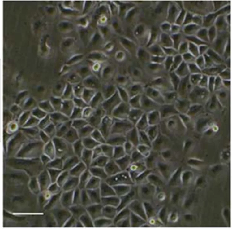
Immortalized Human Epidermal Keratinocytes
Cat.No.: CSC-I9096L
Species: Homo sapiens
Source: Abdominal Skin
Morphology: Polygonal
Culture Properties: Adherent
- Specification
- Background
- Scientific Data
- Q & A
- Customer Review
- Documents
Note: Never can cells be kept at -20 °C.
CIK-HT013 HT® Lenti-hTERT Immortalization Kit
CIK-HT003 HT® Lenti-SV40T Immortalization Kit
Immortalized Human Epidermal Keratinocytes (IHEKs) are human keratinocytes that have been genetically altered to proliferate indefinitely while maintaining important structural and functional features of epidermal basal cells. HaCaT is a spontaneously immortalized human keratinocyte cell line derived from adult human skin and has been well characterized (Boukamp et al., 1988). HaCaT cells are one of the most commonly used immortalized keratinocyte lines.
Under standard growth conditions, IHEKs have a classic cobblestone epithelial morphology and grow as adherent monolayers. They are positive for keratinocyte markers including K5 and K14 (basal state) and can be induced to differentiate with increased calcium concentrations demonstrating upregulation of K1 and K10 as well as involucrin and filaggrin. The ability of immortalized keratinocytes to stratify in an organotypic culture model has also been documented. Overall, these features demonstrate that IHEKs maintain important structural and functional features of normal epidermal cells. IHEKs are commonly used as model systems to study epidermal differentiation and skin barrier formation as well as wound healing, inflammatory signaling (such as activation of NF-κB), UV-induced damage and host-pathogen interactions.
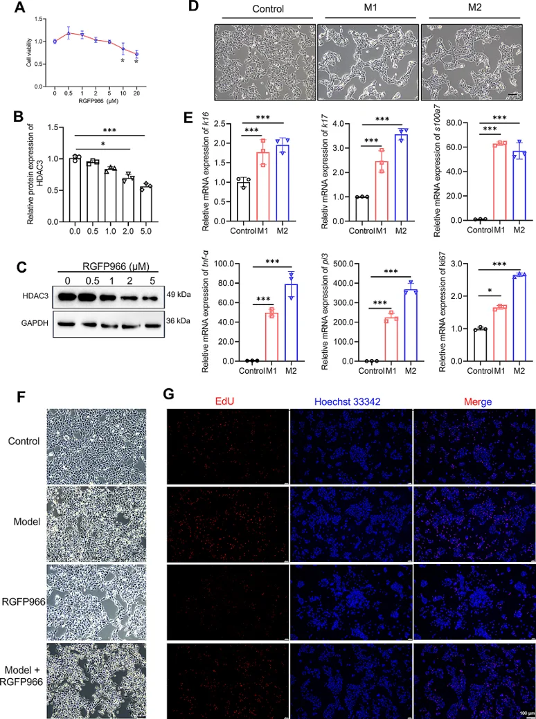
HDAC3 Plays a Critical Role in Keratinocyte Proliferation under Inflammatory Cytokine Stimulation
Psoriasis is known to be mediated by keratinocyte hyperproliferation and immune activation. As HDAC3 is involved in immune regulation and inflammation responses, it is hypothesized that HDAC3 may play a role in psoriasis development. Zeng's team sought to determine HDAC3 expression and contribution towards psoriasis disease models.
Since HDAC3 seemed to contribute to psoriasis phenotype, they tested the functionality of HDAC3 in immortalized human epidermal keratinocytes (HaCaT). CCK8 assays confirmed RGFP966 <10 µM was not cytotoxic to cells (Fig. 1A); they chose 5 µM as their working dose after verifying inhibition of HDAC3 (Figs. 1B-C). Models of psoriasis were created by stimulation of M1 (IL-1α + IL-17A + TNF-α) and M2 (M1 + IL-6) cytokines. Both models showed psoriasis-like morphology and expression of disease-related genes (K16, K17, S100A7, TNF-α, PI3, Ki-67), which was more profound in the M2 model (Fig. 1D-E). Additionally, M2 stimulated cells had increased proliferation that was significantly decreased after treatment with RGFP966 (Fig. 1F-G). These results highlight that HDAC3 contributes to keratinocyte hyperproliferation during psoriatic inflammation and HDAC3 inhibition rescues this disease phenotype.
Ask a Question
Write your own review

Description: Nasal epithelial cells form the outermost protective layer against environmental factors. They clean, humidify, and warm inhaled air. They produces mucus, which binds particles that are subsequently ...
Description: Immortalized Human Corneal Epithelial Cells-SV40 have been obtained immortalizing Human Corneal Epithelial Cells with Lenti-SV40 Lentivirus. Immortalized cells were controlled passaging side by side ...
Description: Immortalized Human Lymphatic Endothelial Cells-SV40 were developed from human tissues transduced with a lentiviral expression vector containing the SV40T gene. The cell line was continuously cultured ...

Description: Immortalized Human Retinal Pigment Epithelial Cells were isolated from neonatal human globes and spontaneously immortalized in culture bypassing crisis. They retained typical morphology of the ...
- Adipose Tissue-Derived Stem Cells
- Human Neurons
- Mouse Probe
- Whole Chromosome Painting Probes
- Hepatic Cells
- Renal Cells
- In Vitro ADME Kits
- Tissue Microarray
- Tissue Blocks
- Tissue Sections
- FFPE Cell Pellet
- Probe
- Centromere Probes
- Telomere Probes
- Satellite Enumeration Probes
- Subtelomere Specific Probes
- Bacterial Probes
- ISH/FISH Probes
- Exosome Isolation Kit
- Human Adult Stem Cells
- Mouse Stem Cells
- iPSCs
- Mouse Embryonic Stem Cells
- iPSC Differentiation Kits
- Mesenchymal Stem Cells
- Immortalized Human Cells
- Immortalized Murine Cells
- Cell Immortalization Kit
- Adipose Cells
- Cardiac Cells
- Dermal Cells
- Epidermal Cells
- Peripheral Blood Mononuclear Cells
- Umbilical Cord Cells
- Monkey Primary Cells
- Mouse Primary Cells
- Breast Tumor Cells
- Colorectal Tumor Cells
- Esophageal Tumor Cells
- Lung Tumor Cells
- Leukemia/Lymphoma/Myeloma Cells
- Ovarian Tumor Cells
- Pancreatic Tumor Cells
- Mouse Tumor Cells