
Canine Dermal Fibroblasts-Adult
Cat.No.: CSC-C4805L
Species: Dog
Source: Dermis; Skin
Cell Type: Fibroblast
- Specification
- Background
- Scientific Data
- Q & A
- Customer Review
Never can cryopreserved cells be kept at -20 °C.
Canine Dermal Fibroblasts (CDFs) are a fibroblastic cell line derived from the dermis of healthy adult dogs (Canis lupus familiaris). As the most abundant stromal cell type in the skin, fibroblasts are major contributors to the synthesis of the ECM and influence the architecture and repair of tissues. As expected of fibroblasts, canine dermal fibroblasts have the spindle-shaped adherent morphology of fibroblasts, and will form aligned bundles of cells, or whorls, upon culture surfaces. Similar to fibroblasts from different species, canine dermal fibroblasts proliferate effectively in DMEM or DMEM/F12 with FBS and demonstrate a doubling time between 24-48 hours but their growth rates show variation depending on the donor source and passage level.
Canine dermal fibroblasts produce various ECM components such as collagens I and III, fibronectin, elastin, and glycosaminoglycans while also responding to multiple cytokines and growth factors. Furthermore, canine dermal fibroblasts can be readily induced to differentiate to α-SMA-positive myofibroblasts in response to TGF-β. As such, they are an ideal cell system to study fibrosis, scarring and contractile wound-healing in vitro. Additionally, canine dermal fibroblasts will produce inflammatory cytokines, chemokines, and other mediators such as IL-6, IL-8, and MIP-1α upon stimulation. This allows CDFs to be used in a variety of dermatologic immunology and infection studies. As CDFs have been isolated directly from canine skin tissue and have not been transformed, they maintain many of the physiologically relevant properties of skin tissue. For this reason, CDFs have been used in many applications including veterinary dermatology, wound healing, toxicology and testing of biomaterials, and mechanistic studies of cellular senescence and cellular stress responses.
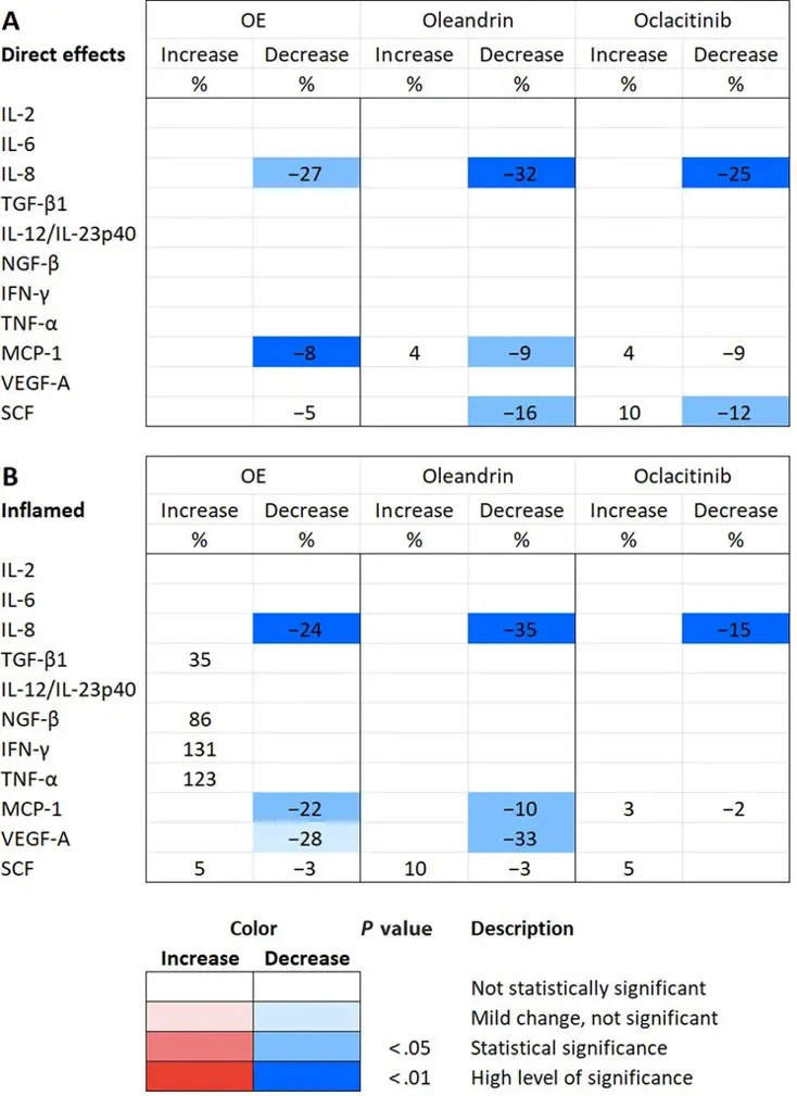
Effects of Botanical Oleander Extract, Oleandrin and Oclacitinib on Canine Dermal Fibroblast Cytokine Secretion
Canine atopic dermatitis involves cytokine-driven dermal inflammation. Here, Fossum et al. tested oleander extract (OE), oleandrin, and oclacitinib on primary canine dermal fibroblasts and DH82 macrophages under inflamed conditions.
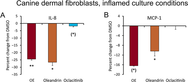
Cytokine levels in supernatants from canine dermal fibroblasts were measured using a canine 11-plex detection kit. Detectable cytokines included IL-6, IL-8, MCP-1 (CCL2), VEGF-A, and SCF. As shown (Fig. 1), all three test products reduced IL-8, MCP-1, and SCF levels. For IL-8, reductions were similar across products. For MCP-1, OE caused the most significant reduction, while for SCF, oleandrin had the greatest effect and OE the mildest. Under LPS-induced inflammation, all products reduced IL-8 secretion, with OE and oleandrin showing robust effects and oclacitinib a mild effect (Fig. 1). VEGF-A levels were significantly reduced by OE and oleandrin, with oleandrin showing statistical significance for both cytokines and OE for MCP-1. Oclacitinib did not affect MCP-1 levels, while OE and oleandrin significantly reduced its secretion (Fig. 2). OE and oleandrin showed anti-inflammatory effects on IL-8 and MCP-1 at a low dose of 0.03 μg/mL, whereas oclacitinib at 0.01 μM was not effective in reducing these cytokines.
Ask a Question
Write your own review
Description: Dog Liver Endothelial Cells from Creative Bioarray are isolated from tissue of dog liver. Dog Liver Endothelial Cells are grown in T25 tissue culture flasks pre-coated with gelatin-based coating ...
Description: Canine Astrocytes from Creative Bioarray are isolated from canine brain tissue. The method we use to isolate canine astrocytes were developed based on a combination of established and our proprietary ...
Description: Canine Mammary Microvascular Endothelial Cells from Creative Bioarray are isolated from breast of pathogen-free laboratory Canine. Canine Mammary Microvascular Endothelial Cells are grown in T25 ...
Description: Canine Chondrocytes (CnC) provided by Creative Bioarray are isolated from normal canine articular cartilage tissue. The cells are frozen at passage 1 and each vial contains at least 0.5*10^6 cells. ...
Description: Canine Pancreatic Microvascular Endothelial Cells from Creative Bioarray are isolated from Pancreatic Microvascular of pathogen-free laboratory Canine. Canine Pancreatic Microvascular Endothelial ...
Description: Canine Prostate Microvascular Endothelial Cells from Creative Bioarray are isolated from prostate of pathogen-free laboratory Canine. Canine Prostate Microvascular Endothelial Cells are grown in T25 ...