
Human Gastric Smooth Muscle Cells (HGSMC)
Cat.No.: CSC-7779W
Species: Human
Source: Stomach
Cell Type: Smooth Muscle Cell
- Specification
- Background
- Scientific Data
- Q & A
- Customer Review
Human gastric smooth muscle cells (HGSMC) are primary cells isolated from human stomach tissue that provide a physiologically relevant in vitro model system for studying gastric motility as well as other smooth muscle-mediated gastrointestinal functions. HGSMCs maintain many structural, molecular, and functional properties of gastric smooth muscle cells in vivo. HGSMCs are spindle-shaped elongated cells that attach and proliferate as monolayers under conventional culture conditions. They also express several smooth muscle specific proteins such as α-smooth muscle actin (α-SMA), smooth muscle myosin heavy chain, calponin, and desmin. Functionally, HGSMCs contract and relax in response to neural and hormonal regulators of gastric motility, including acetylcholine (muscarinic receptors), nitric oxide-dependent relaxation pathways, and calcium-induced contraction.
HGSMCs are commonly utilized to explore basic mechanisms underlying diseases associated with gastric motility dysfunction, such as gastroparesis, functional dyspepsia, and inflammatory-induced gastric smooth muscle cell dysfunction. Furthermore, they serve as an excellent cell model for screening prokinetic/antispasmodic drug candidates, as well as studying smooth muscle proliferation, phenotype switching, and extracellular matrix remodeling.
Maslinic Acid Improves Mitochondrial Function and Inhibits Oxidative Stress and Autophagy in Human Gastric Smooth Muscle Cells
Functional dyspepsia (FD) is a common chronic gastroduodenal disorder lacking efficient pharmacotherapy. Zheng's team explored if maslinic acid (MA), pentacyclic triterpene acid extracted from hawthorn, exerted cytoprotection on human gastric smooth-muscle cells (HGSMCs) against mitochondrial damage and further investigated the mechanisms involved.
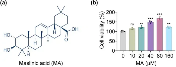
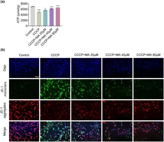
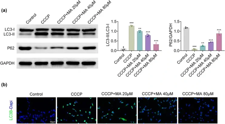
Figure 1a displayed the chemical structure of MA. After that, we found that cell viability was progressively augmented after treatment with MA (20, 40, 80 and 160 μM), and then 20, 40 and 80 μM MA treatments were selected for subsequent studies (Fig. 1b). Taken together, MA promoted cell viability in HGSMCs. They found that the ATP level was decreased after CCCP induction while this decrease was abrogated after MA treatment (20, 40 and 80 μM) (Fig. 2a). Additionally, the MMP levels decreased after CCCP addition, but this effect was reversed after MA treatment (Fig. 2b). Taken together, MA ameliorated mitochondrial function in HGSMCs. The protein expression of LC3II/LC3I was increased and that of P62 was attenuated after CCCP stimulation, but these impacts were counteracted after MA addition (Fig. 3a). Moreover, the LC3B fluorescence intensity was heightened after CCCP induction, but this increase was neutralized after MA treatment (Fig. 3a and b). Overall, MA restrained autophagy in HGSMCs.
Ask a Question
Write your own review
Description: Fibroblasts are mesenchymal cells derived from the embryonic mesoderm. They have been extensively used for a wide range of cellular and molecular studies. This is mainly because they are one of ...

Description: Smooth muscle contraction is the fundamental event in gastrointestinal motion. Inflammation of the human intestine causes thickening of the smooth muscle layers which results from the increases in ...
Description: The colorectum is a major organ for both malignant and nonmalignant diseases. Cells that line the colonic mucosal surface form a major mechanical barrier that separates the host's internal milieu ...
Description: Primary Human Large Intestine Microvascular Endothelial Cells were initiated from normal human large intestine (colon) tissue.These cells were originated using Complete Serum-Free Medium Kit With ...
Description: Human Primary Intestinal Vascular Fibroblasts from Creative Bioarray are isolated from normal human intestinal tissue. Human Primary Intestinal Vascular Fibroblasts are grown in T75 tissue culture ...
Description: Human Small Intestinal Microvascular Endothelial Cells from Creative Bioarray are isolated from human small intestinal tissue. Human Small Intestinal Microvascular Endothelial Cells are grown in T25 ...