

Immortalized Human Melanocytes-hTERT
Cat.No.: CSC-I9169L
Species: Homo sapiens
Source: Skin
Morphology: Polygonal
Culture Properties: Adherent
- Specification
- Background
- Scientific Data
- Q & A
- Customer Review
- Documents
Note: Never can cells be kept at -20 °C.
Immortalized Human Melanocytes-hTERT are normal human melanocytes that have been stably immortalized by the ectopic expression of human telomerase reverse transcriptase (hTERT). Originating from primary human epidermal or dermal melanocytes, these cells circumvent replicative senescence by continuous telomere preservation, enabling prolonged and stable in vitro passage while maintaining a nearly normal diploid karyotype and non-tumorigenic characteristics. They have characteristic dendritic architecture and express essential melanocytic markers, including tyrosinase (TYR), TRP‑1, TRP‑2, MITF, S100, and HMB45, while maintaining physiological processes like as melanin production and melanosome formation. In contrast to primary melanocytes, hTERT-immortalized melanocytes exhibit less batch variability and a longer culture lifetime. This cell line functions as a dependable in vitro model extensively utilized in the investigation of melanocyte biology, pigmentation control, skin photodamage, screening of cosmetic whitening agents, and the initial causes of melanocytic diseases.
Syntenin Regulates Melanogenesis via the P38 MAPK Pathway
Melanogenesis, the production of melanin, is important in studying pigmentary skin illnesses. Syntenin is a melanosome protein, however its involvement in melanogenesis is unclear. Sun's team examined syntenin's effects and mechanisms on immortalized human melanocyte melanogenesis.
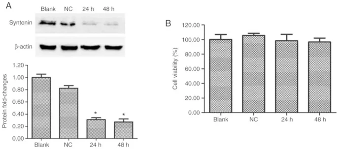
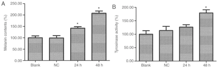
Western blot analysis was conducted to test if syntenin siRNA transfections decreased PIG1 cell syntenin protein expression. Syntenin-siRNA-transfected melanocytes had lower syntenin protein expression than NC-siRNA-transfected melanocytes at 24 and 48 h after transfection (Fig. 1A). Over 48 h, syntenin-siRNA transfection did not affect cell viability (Fig. 1B). These data showed that syntenin-siRNA transfection decreased syntenin. Melanin and Tyr enzyme activity were measured 24 and 48 h following syntenin-siRNA transfection of PIG1 cells. Compared to NC-siRNA-transfected cells, syntenin-siRNA-transfected cells had more melanin and Tyr activity (Fig. 2A and B). In immortalized human melanocytes, syntenin deficiency caused melanogenesis.
Ask a Question
Write your own review
 Immortalized Human Melanocytes-hTERT
Description: Nasal epithelial cells form the outermost protective layer against environmental factors. They clean, humidify, and warm inhaled air. They produces mucus, which binds particles that are subsequently ...
Description: Immortalized Human Splenic Endothelial Cells-SV40 have been obtained immortalizing Human Splenic Endothelial Cells with SV40LT expressing lentiviral particles. Immortalized cells were controlled ...
Description: Immortalized Human Corneal Epithelial Cells-SV40 have been obtained immortalizing Human Corneal Epithelial Cells with Lenti-SV40 Lentivirus. Immortalized cells were controlled passaging side by side ...
Description: Immortalized Human Lymphatic Endothelial Cells-SV40 were developed from human tissues transduced with a lentiviral expression vector containing the SV40T gene. The cell line was continuously cultured ...
- Adipose Tissue-Derived Stem Cells
- Human Neurons
- Mouse Probe
- Whole Chromosome Painting Probes
- Hepatic Cells
- Renal Cells
- In Vitro ADME Kits
- Tissue Microarray
- Tissue Blocks
- Tissue Sections
- FFPE Cell Pellet
- Probe
- Centromere Probes
- Telomere Probes
- Satellite Enumeration Probes
- Subtelomere Specific Probes
- Bacterial Probes
- ISH/FISH Probes
- Exosome Isolation Kit
- Human Adult Stem Cells
- Mouse Stem Cells
- iPSCs
- Mouse Embryonic Stem Cells
- iPSC Differentiation Kits
- Mesenchymal Stem Cells
- Immortalized Human Cells
- Immortalized Murine Cells
- Cell Immortalization Kit
- Adipose Cells
- Cardiac Cells
- Dermal Cells
- Epidermal Cells
- Peripheral Blood Mononuclear Cells
- Umbilical Cord Cells
- Monkey Primary Cells
- Mouse Primary Cells
- Breast Tumor Cells
- Colorectal Tumor Cells
- Esophageal Tumor Cells
- Lung Tumor Cells
- Leukemia/Lymphoma/Myeloma Cells
- Ovarian Tumor Cells
- Pancreatic Tumor Cells
- Mouse Tumor Cells