

Immortalized Human Lymphoblastoid Cells-EBV (KMS-15)
Cat.No.: CSC-I9250L
Species: Homo sapiens
Source: Peripheral blood
Morphology: Lymphoblast-like
Culture Properties: Suspension
- Specification
- Background
- Scientific Data
- Q & A
- Customer Review
The KMS-15 cells are positive for both CD19 and CD20 B cell antigens as well as HLA-DR. In contrast, another lyphoblastoid cell line, KMS-9 is positive for HLA-DR and CD19B while tested negative for CD20.
Note: Never can cells be kept at -20 °C.
Immortalized Human Lymphoblastoid Cells-EBV are human B lymphocyte cell lines generated by infection with Epstein-Barr virus (EBV) that results in cellular immortalization. Upon EBV-mediated transformation, quiescent peripheral B cells acquire infinite proliferative ability while retaining numerous phenotypic, genomic and immunological features of their donor origin. These lymphoblastoid cell lines (LCLs) are among the most extensively used human cellular models in immunology, genetics and biomedical research.
Immortalized Human Lymphoblastoid Cells are normally cultured in suspension, can be expanded stably over lengthy periods of time and produce B-cell markers, major histocompatibility complex (MHC) molecules and EBV latent gene products. They are excellent renewable sources of human genomic material, and are widely used in studies of host-virus interactions, B-cell biology, antigen presentation, immunological modulation and Epstein-Barr virus-associated illnesses owing to their genetic stability and simplicity of culture.
These cells have been widely used for pharmacogenomics, functional genomics, gene expression profiling, epigenetic analysis, genome-wide association studies (GWAS) and personalized medicine research. They also provide powerful experimental models for studying viral oncogenesis, lymphoproliferative diseases, immunological signaling pathways and identification of therapeutic targets. The Immortalized Human Lymphoblastoid Cells–EBV line, a well-established and highly reproducible human cell model, still plays a crucial role in the advancement of translational research and precision medicine.
Long-term Culture of EBV-induced Human Lymphoblastoid Cell Lines Reveals Chromosomal Instability
Lymphoblastoid cell lines (LCLs) are essential for genetic research, yet their genomic stability during prolonged culture is poorly defined. Volleth et al. performed serial cytogenetic analyses at 8-week intervals for up to three years across 10 LCLs derived from individuals with normal, marker-bearing, or aberrant baseline karyotypes.
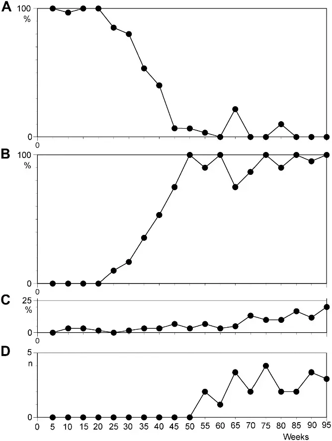
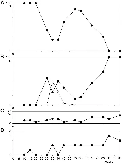
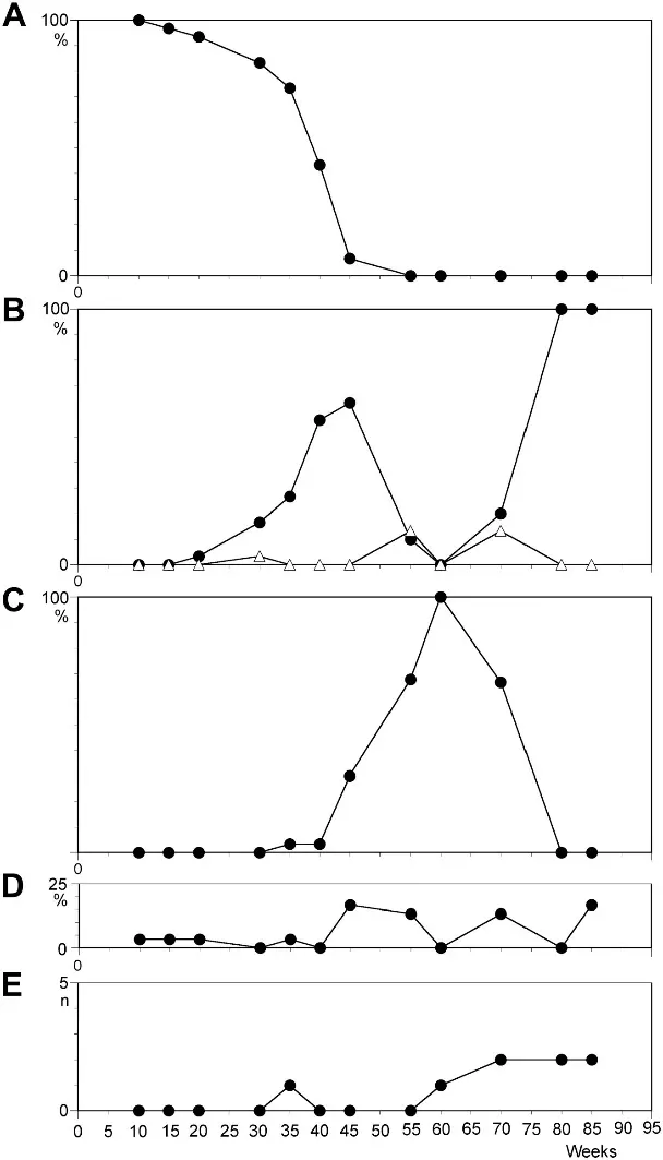
All LCLs maintained their original karyotype only during the initial months of culture. Subsequently, every cell line acquired clonal chromosomal aberrations, with the proportion of cytogenetically normal cells declining continuously over time. Notably, karyotype evolution varied even between lines from the same donor. The most prevalent abnormality was trisomy 12, emerging as a clonal event in all cell lines between 21–49 weeks post-infection (median: 27 weeks). This clone expanded rapidly, achieving >50% prevalence within 27–62 weeks (median: 39.5 weeks) and requiring a median of only 20.5 weeks to progress from undetectable to dominant (>50% of metaphases). By the end of the observation period, seven lines harbored trisomy 12 in >90% of cells (Fig. 1). One line progressed to hypertetraploidy (50 chromosomes; +5, +10, +12, +12) before dying, while another acquired structural rearrangements alongside trisomy 12. The results for cell lines N1 and N2 are shown in Figs. 2 and 3, respectively. Unexpectedly, one line reverted to its baseline karyotype despite prior trisomy 12 dominance. Three lines ceased proliferating during monitoring; all had acquired clonal aberrations beforehand, demonstrating that chromosomal instability does not preclude crisis or guarantee immortalization. These findings establish trisomy 12 as a near-universal, early, and dominant event in LCL evolution, underscoring the necessity of cryopreserving early-passage aliquots to preserve the donor's primary genetic architecture.
Ask a Question
Write your own review

Description: Nasal epithelial cells form the outermost protective layer against environmental factors. They clean, humidify, and warm inhaled air. They produces mucus, which binds particles that are subsequently ...
Description: Immortalized Human Splenic Endothelial Cells-SV40 have been obtained immortalizing Human Splenic Endothelial Cells with SV40LT expressing lentiviral particles. Immortalized cells were controlled ...
Description: Immortalized Human Corneal Epithelial Cells-SV40 have been obtained immortalizing Human Corneal Epithelial Cells with Lenti-SV40 Lentivirus. Immortalized cells were controlled passaging side by side ...
Description: Immortalized Human Lymphatic Endothelial Cells-SV40 were developed from human tissues transduced with a lentiviral expression vector containing the SV40T gene. The cell line was continuously cultured ...

- Adipose Tissue-Derived Stem Cells
- Human Neurons
- Mouse Probe
- Whole Chromosome Painting Probes
- Hepatic Cells
- Renal Cells
- In Vitro ADME Kits
- Tissue Microarray
- Tissue Blocks
- Tissue Sections
- FFPE Cell Pellet
- Probe
- Centromere Probes
- Telomere Probes
- Satellite Enumeration Probes
- Subtelomere Specific Probes
- Bacterial Probes
- ISH/FISH Probes
- Exosome Isolation Kit
- Human Adult Stem Cells
- Mouse Stem Cells
- iPSCs
- Mouse Embryonic Stem Cells
- iPSC Differentiation Kits
- Mesenchymal Stem Cells
- Immortalized Human Cells
- Immortalized Murine Cells
- Cell Immortalization Kit
- Adipose Cells
- Cardiac Cells
- Dermal Cells
- Epidermal Cells
- Peripheral Blood Mononuclear Cells
- Umbilical Cord Cells
- Monkey Primary Cells
- Mouse Primary Cells
- Breast Tumor Cells
- Colorectal Tumor Cells
- Esophageal Tumor Cells
- Lung Tumor Cells
- Leukemia/Lymphoma/Myeloma Cells
- Ovarian Tumor Cells
- Pancreatic Tumor Cells
- Mouse Tumor Cells