

Immortalized Human Kidney Podocytes-SV40
Cat.No.: CSC-I2122Z
Species: homo sapiens
Morphology: Polygonal
Culture Properties: Adherent
- Specification
- Background
- Scientific Data
- Q & A
- Customer Review
free from contaminations (bacteria incl. mycoplasma, fungi, HIV, HAV, HBV, HCV, Parvo-B19) and cross-contaminations
Note: Never can cells be kept at -20°C.
The Immortalized Human Kidney Podocytes-SV40 bypass replicative senescence through inactivation of p53 and pRB pathways, thereby acquiring continuous proliferation while preserving their differentiated phenotype under permissive culture conditions (e.g., 33 °C with interferon-γ), and can be induced to differentiate at 37 °C.
Key advantages of this cell line include: (1) maintenance of characteristic podocyte morphology, including arborized processes with primary and secondary branches; (2) expression of signature proteins such as nephrin, podocin, synaptopodin, WT-1, and CD2AP, confirmed by immunoblotting and immunofluorescence; (3) formation of rudimentary slit diaphragm-like structures; (4) functional responsiveness to mechanical and pharmacological stimuli, including cytoskeletal reorganization upon puromycin aminonucleoside or angiotensin II exposure; and (5) long-term genetic stability enabling consistent experimental outcomes.
Collectively, Immortalized Human Kidney Podocytes-SV40 provide an unlimited, phenotypically faithful, and experimentally tractable in vitro model for investigating podocyte biology, glomerular diseases (e.g., focal segmental glomerulosclerosis, diabetic nephropathy), and nephrotoxic drug screening, circumventing the inherent limitations of primary cultures.
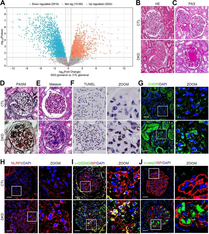
Unfolded Protein Response-Activated NLRP3 Inflammasome Contributes To Pyroptotic and Apoptotic Podocyte Injury in Diabetic Kidney Disease
Diabetic kidney disease (DKD) is the leading cause of chronic kidney disease and end-stage renal disease worldwide. Podocyte injury and death is a key event in DKD progression. Emerging evidence has indicated that crosstalk between unfolded protein response (UPR) and NLR family pyrin domain containing 3 (NLRP3) inflammasome plays an essential role in DKD progression. However, the involvement of these pathways in podocyte injury and death during DKD remains unclear.
Here, we found that inositol-requiring enzyme 1 (IRE1) and protein kinase RNA-like ER kinase (PERK) branches of the UPR, NLRP3 inflammasome, and apoptosis were activated in podocytes under DKD and high glucose (HG) conditions. In vitro, inducing ER stress by thapsigargin, and IRE1 or PERK overexpression upon HG treatment stimulated NLRP3 inflammasome-mediated pyroptosis and apoptosis, whereas inhibiting IRE1 or PERK suppressed them. Importantly, we discovered that the newly identified NLRP3-binding partner, thioredoxin-interacting protein (TXNIP), upon activation by the transcription factor (TF) PERK/CCAAT-enhancer-binding protein homologous protein (CHOP), served as a link between IRE1 or PERK branches with NLRP3 inflammasome-mediated pyroptosis and apoptosis.
In conclusion, the results suggested that UPR/NLRP3 inflammasome-mediated pyroptosis/apoptosis pathway was involved in diabetic podocyte injury, and that targeting the CHOP-TXNIP axis may serve as a promising therapeutic target for DKD.
Ask a Question
Write your own review
Description: Nasal epithelial cells form the outermost protective layer against environmental factors. They clean, humidify, and warm inhaled air. They produces mucus, which binds particles that are subsequently ...

Description: Immortalized Human Splenic Endothelial Cells-SV40 have been obtained immortalizing Human Splenic Endothelial Cells with SV40LT expressing lentiviral particles. Immortalized cells were controlled ...
Description: Immortalized Human Corneal Epithelial Cells-SV40 have been obtained immortalizing Human Corneal Epithelial Cells with Lenti-SV40 Lentivirus. Immortalized cells were controlled passaging side by side ...
Description: Immortalized Human Lymphatic Endothelial Cells-SV40 were developed from human tissues transduced with a lentiviral expression vector containing the SV40T gene. The cell line was continuously cultured ...
- Adipose Tissue-Derived Stem Cells
- Human Neurons
- Mouse Probe
- Whole Chromosome Painting Probes
- Hepatic Cells
- Renal Cells
- In Vitro ADME Kits
- Tissue Microarray
- Tissue Blocks
- Tissue Sections
- FFPE Cell Pellet
- Probe
- Centromere Probes
- Telomere Probes
- Satellite Enumeration Probes
- Subtelomere Specific Probes
- Bacterial Probes
- ISH/FISH Probes
- Exosome Isolation Kit
- Human Adult Stem Cells
- Mouse Stem Cells
- iPSCs
- Mouse Embryonic Stem Cells
- iPSC Differentiation Kits
- Mesenchymal Stem Cells
- Immortalized Human Cells
- Immortalized Murine Cells
- Cell Immortalization Kit
- Adipose Cells
- Cardiac Cells
- Dermal Cells
- Epidermal Cells
- Peripheral Blood Mononuclear Cells
- Umbilical Cord Cells
- Monkey Primary Cells
- Mouse Primary Cells
- Breast Tumor Cells
- Colorectal Tumor Cells
- Esophageal Tumor Cells
- Lung Tumor Cells
- Leukemia/Lymphoma/Myeloma Cells
- Ovarian Tumor Cells
- Pancreatic Tumor Cells
- Mouse Tumor Cells