
Immortalized Human Kidney Fibroblasts-SV40
Cat.No.: CSC-I2121Z
Species: homo sapiens
Morphology: Polygonal
Culture Properties: Adherent
- Specification
- Background
- Scientific Data
- Q & A
- Customer Review
free from contaminations (bacteria incl. mycoplasma, fungi, HIV, HAV, HBV, HCV, Parvo-B19) and cross-contaminations
Note: Never can cells be kept at -20°C.
Human Kidney Fibroblasts-SV40 Immortalized are human renal fibroblasts that have been transfected with the SV40 Large T antigen to achieve extended proliferation while retaining critical fibroblast properties. These cells provide a stable and reproducible in vitro model for the investigation of kidney fibrosis, extracellular matrix (ECM) remodeling and renal microenvironment interactions. SV40-immortalized cells are excellent for long-term mechanistic and pharmacological research, as they are free from the limits of donor variability, short lifespan and early senescence associated with primary kidney fibroblasts.
Immortalized Human Kidney Fibroblasts-SV40 often retain the fibroblast spindle-shaped morphology and express fibroblast-associated markers involved in matrix formation and fibrotic signaling pathways. They are commonly employed in the research of chronic kidney disease (CKD), renal inflammation, epithelial-mesenchymal transition (EMT), and TGF-β/Smad-mediated fibrosis. These cells are also useful for cell-cell communication studies in the renal microenvironment and for antifibrotic drug candidate screening.
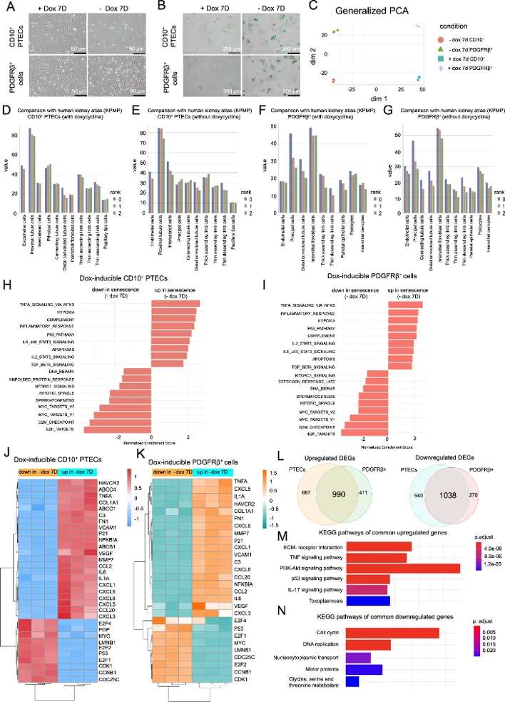
Bulk Transcriptome Analysis of Conditionally Immortalized Human Kidney CD10+ PTECs and PDGFRΒ+ Fibroblasts
Cellular senescence is a major contributor to the pathogenesis of chronic kidney disease (CKD) especially in proximal tubule epithelial cells (PTECs) and fibroblasts. Current cellular models are often confounded by age and comorbidities and human kidney cell lines often poorly represent in vivo physiology due to cancer-like changes. The researchers aimed to establish a controllable conditionally immortalized human kidney cell model (doxycycline inducible SV40LT) for the purpose of accurately mimicking in vivo cellular behavior, mechanistic exploration of senescence-specific processes, and verifying clinical relevance of senescence in CKD progression through ex vivo and in vivo correlations.
They performed bulk RNAseq to characterize transcriptional changes upon doxycycline withdrawal that induces cellular senescence. Principal component analysis (PCA) showed separation by cell identity (epithelial vs mesenchymal) and senescence status (Fig. 1C). Validation with single-cell data from Kidney Precision Medicine Project (KPMP) supported our CD10+ PTECs and PDGFRβ+ The fibroblasts were very much like the in vivo cells, proximal tubule cells and interstitial fibroblasts respectively (Fig. 1D-G).
Senescent cells exhibited hallmark features such as increased NF-κB, inflammatory response and p53 signaling, while pathways of cell proliferation were decreased (Fig. 1H, I). Key markers of senescence (CDKN1A, LMNB1) and genes involved in inflammation, fibrosis and chemotaxis were concordantly changed in both cell lines (Fig. 1J, K). KEGG analysis further confirmed this common senescence signature by showing activated TNF-α/p53 signaling pathway and inhibited cell cycle pathways (Fig. 1L-N).
Ask a Question
Write your own review

Description: Nasal epithelial cells form the outermost protective layer against environmental factors. They clean, humidify, and warm inhaled air. They produces mucus, which binds particles that are subsequently ...
Description: Immortalized Human Splenic Endothelial Cells-SV40 have been obtained immortalizing Human Splenic Endothelial Cells with SV40LT expressing lentiviral particles. Immortalized cells were controlled ...
Description: Immortalized Human Corneal Epithelial Cells-SV40 have been obtained immortalizing Human Corneal Epithelial Cells with Lenti-SV40 Lentivirus. Immortalized cells were controlled passaging side by side ...
Description: Immortalized Human Lymphatic Endothelial Cells-SV40 were developed from human tissues transduced with a lentiviral expression vector containing the SV40T gene. The cell line was continuously cultured ...
- Adipose Tissue-Derived Stem Cells
- Human Neurons
- Mouse Probe
- Whole Chromosome Painting Probes
- Hepatic Cells
- Renal Cells
- In Vitro ADME Kits
- Tissue Microarray
- Tissue Blocks
- Tissue Sections
- FFPE Cell Pellet
- Probe
- Centromere Probes
- Telomere Probes
- Satellite Enumeration Probes
- Subtelomere Specific Probes
- Bacterial Probes
- ISH/FISH Probes
- Exosome Isolation Kit
- Human Adult Stem Cells
- Mouse Stem Cells
- iPSCs
- Mouse Embryonic Stem Cells
- iPSC Differentiation Kits
- Mesenchymal Stem Cells
- Immortalized Human Cells
- Immortalized Murine Cells
- Cell Immortalization Kit
- Adipose Cells
- Cardiac Cells
- Dermal Cells
- Epidermal Cells
- Peripheral Blood Mononuclear Cells
- Umbilical Cord Cells
- Monkey Primary Cells
- Mouse Primary Cells
- Breast Tumor Cells
- Colorectal Tumor Cells
- Esophageal Tumor Cells
- Lung Tumor Cells
- Leukemia/Lymphoma/Myeloma Cells
- Ovarian Tumor Cells
- Pancreatic Tumor Cells
- Mouse Tumor Cells