

Immortalized Human Hepatic Stellate Cells-SV40 (Tet-on)
Cat.No.: CSC-I1913Z
Species: homo sapiens
Morphology: Polygonal
Culture Properties: Adherent
- Specification
- Background
- Scientific Data
- Q & A
- Customer Review
CIK-HT003 HT? Lenti-SV40T Immortalization Kit
CIK-HT004 HT? Lenti-SV40 (tsA58 temperature sensitive mutant) Lentivirus Immortalization Kit
Note: Never can cells be kept at -20 °C.
Immortalized Human Hepatic Stellate Cells-SV40 (Tet-on) are human hepatic stellate cells (HSC) that have been immortalized via lentiviral transduction of the SV40 large T antigen, controlled by a doxycycline-inducible Tet-on promoter. As a result of this controlled induction system these cells can be conditionally immortalized while retaining normal cellular function. This cell line is adherent, displays normal stellate cell morphology and can be maintained for long-term culture and repeated experiments.
Hepatic stellate cells are major components of non-parenchymal cells in the liver and line the space of Disse. HSCs play an important role in the production and regulation of extracellular matrix (ECM). In response to liver damage or injury HSCs can undergo activation and are responsible for the majority of collagen and pro-fibrotic cytokine production leading to fibrosis. SV40-immortalized HSC models maintain important phenotypic characteristics such as expression of α-SMA, collagen I, and activation by fibrogenic mediators such as TGF-β. This allows for detailed mechanistic studies into fibrosis and ECM regulation. Addition of Tet-on system allows for additional gene induction as needed for experimental flexibility as well as reducing potential effects of constitutive oncogene expression.
Applications of these cells include basic research into liver fibrosis, high throughput screening of anti-fibrotic drugs and co-culture with hepatocytes for bioartificial liver purposes. Immortalized Human Hepatic Stellate Cells-SV40 (Tet-on) allow for controlled, reproducible and physiologically relevant in vitro experimentation.
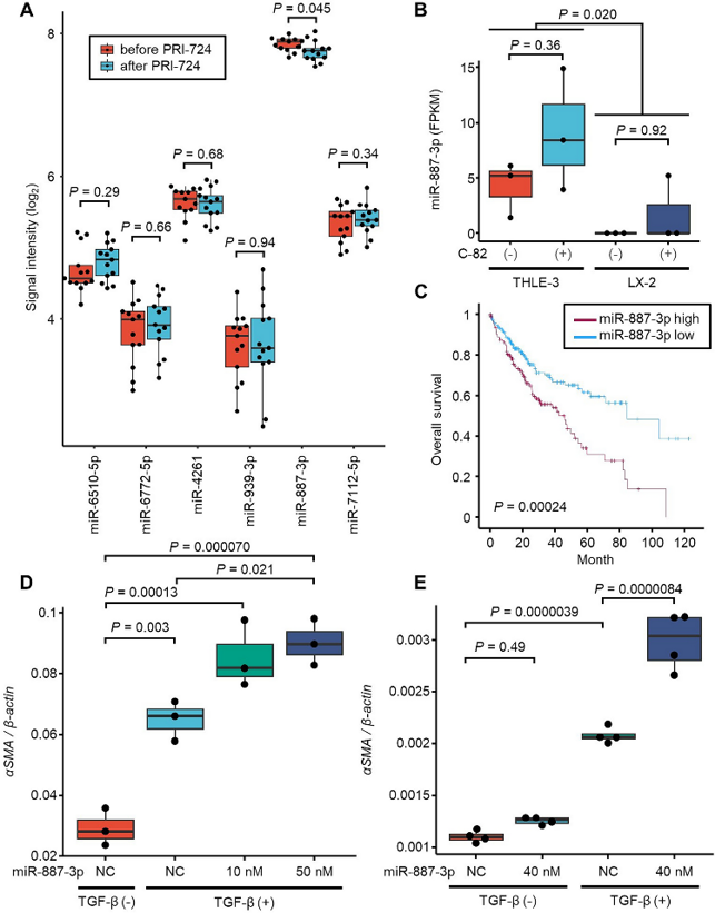
Hepatocyte‑Derived miR‑887‑3p as a Response Biomarker and Independent Prognostic Factor in PRI‑724 Antifibrotic Therapy
There are currently no antifibrotic therapies for liver cirrhosis. Yoshida et al. previously conducted a phase I trial demonstrating the safety and potential antifibrotic activity of the investigational agent PRI-724. They sought to determine which circulating EV-miRNAs were associated with treatment response and which were associated with treatment efficacy after therapy with PRI-724 as potential biomarkers for antifibrotic therapies.
Three miRNAs were associated with response to PRI-724 (miR-6510-5p, miR-6772-5p, miR-4261), and three were associated with treatment efficacy (miR-939-3p, miR-887-3p, miR-7112-5p). Of these six miRNAs tested in liver tissue, only miR-887-3p had a signal intensity >26 and significantly changed levels after PRI-724 treatment (P=0.045) (Fig. 1A).
ISH demonstrated that miR-887-3p was expressed in hepatocytes similarly to the hepatocyte enriched miR-122-3p. qPCR of EV-miRNAs from immortalized hepatocytes (THLE-3) and hepatic stellate cells (LX-2) revealed miR-887-3p expression only in THLE-3 immortalized hepatocytes suggesting hepatocytes are the primary source of miR-887-3p. Exposure to C-82, the active metabolite of PRI-724 did not significantly change expression of miR-887-3p in THLE-3 cells(Fig. 1B).
Of interest, increased expression of miR-887-3p in HCC tissue was associated with worse prognosis using the TCGA-LIHC dataset (Fig. 1C). Tissue miR-887-3p was also determined to be an independent prognostic factor for HCC through Cox regression. These data suggest inhibition of miR-887-3p may provide cytoprotective effects to the liver.
Ask a Question
Write your own review

Description: Nasal epithelial cells form the outermost protective layer against environmental factors. They clean, humidify, and warm inhaled air. They produces mucus, which binds particles that are subsequently ...

Description: Immortalized Human Splenic Endothelial Cells-SV40 have been obtained immortalizing Human Splenic Endothelial Cells with SV40LT expressing lentiviral particles. Immortalized cells were controlled ...
Description: Immortalized Human Corneal Epithelial Cells-SV40 have been obtained immortalizing Human Corneal Epithelial Cells with Lenti-SV40 Lentivirus. Immortalized cells were controlled passaging side by side ...
Description: Immortalized Human Lymphatic Endothelial Cells-SV40 were developed from human tissues transduced with a lentiviral expression vector containing the SV40T gene. The cell line was continuously cultured ...
- Adipose Tissue-Derived Stem Cells
- Human Neurons
- Mouse Probe
- Whole Chromosome Painting Probes
- Hepatic Cells
- Renal Cells
- In Vitro ADME Kits
- Tissue Microarray
- Tissue Blocks
- Tissue Sections
- FFPE Cell Pellet
- Probe
- Centromere Probes
- Telomere Probes
- Satellite Enumeration Probes
- Subtelomere Specific Probes
- Bacterial Probes
- ISH/FISH Probes
- Exosome Isolation Kit
- Human Adult Stem Cells
- Mouse Stem Cells
- iPSCs
- Mouse Embryonic Stem Cells
- iPSC Differentiation Kits
- Mesenchymal Stem Cells
- Immortalized Human Cells
- Immortalized Murine Cells
- Cell Immortalization Kit
- Adipose Cells
- Cardiac Cells
- Dermal Cells
- Epidermal Cells
- Peripheral Blood Mononuclear Cells
- Umbilical Cord Cells
- Monkey Primary Cells
- Mouse Primary Cells
- Breast Tumor Cells
- Colorectal Tumor Cells
- Esophageal Tumor Cells
- Lung Tumor Cells
- Leukemia/Lymphoma/Myeloma Cells
- Ovarian Tumor Cells
- Pancreatic Tumor Cells
- Mouse Tumor Cells