

Immortalized Human Gingival Keratinocytes-hTERT
Cat.No.: CSC-I2300Z
Morphology: Polygonal
Culture Properties: Adherent
- Specification
- Background
- Scientific Data
- Q & A
- Customer Review
Immortalized human gingival keratinocytes-hTERT are established by transducing primary human gingival keratinocytes with a lentiviral expression vector encoding the human telomerase reverse transcriptase (hTERT) gene. The advantages of hTERT-immortalized gingival keratinocytes are threefold. First, extended proliferative capacity – whereas primary cells senesce within a few passages, hTERT-transduced cells can be continuously cultured beyond 20 passages without growth retardation or replicative senescence. The telomerase immortalized gingival keratinocyte (TIGK) line has been reported to undergo 36 passages while maintaining robust proliferation. Second, phenotypic fidelity – immortalized cells retain morphological characteristics, karyotypic stability, proliferation rates, and cytokeratin expression profiles (including cytokeratins 2, 17 and 19) comparable to primary gingival keratinocytes. When cultured under stratifying conditions, they recapitulate suprabasal differentiation markers consistent with junctional epithelium. This preservation of lineage-specific traits distinguishes hTERT-immortalized lines from virally immortalized alternatives, which may exhibit chromosomal rearrangements or aberrant differentiation. Third, experimental reproducibility and scalability – the consistent growth and stable phenotype eliminate the donor-to-donor variability inherent to primary cultures, enabling repeated experiments and high-throughput screening. These cells are amenable to co-culture with fibroblasts for constructing three-dimensional gingival tissue equivalents and organotypic multilayered models, and have been successfully integrated into full-thickness reconstructed human gingiva for wound healing and toxicological studies. Collectively, hTERT-immortalized human gingival keratinocytes offer a robust, phenotypically faithful, and ethically sustainable in vitro model system that bridges the gap between primary cell biology and translational oral research.
Differential Temperature-Induced Responses in Immortalized Oral and Skin Keratinocytes
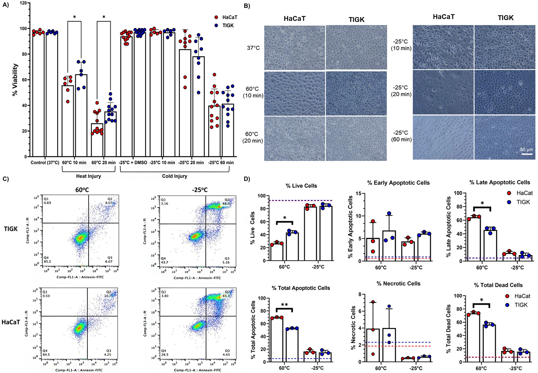
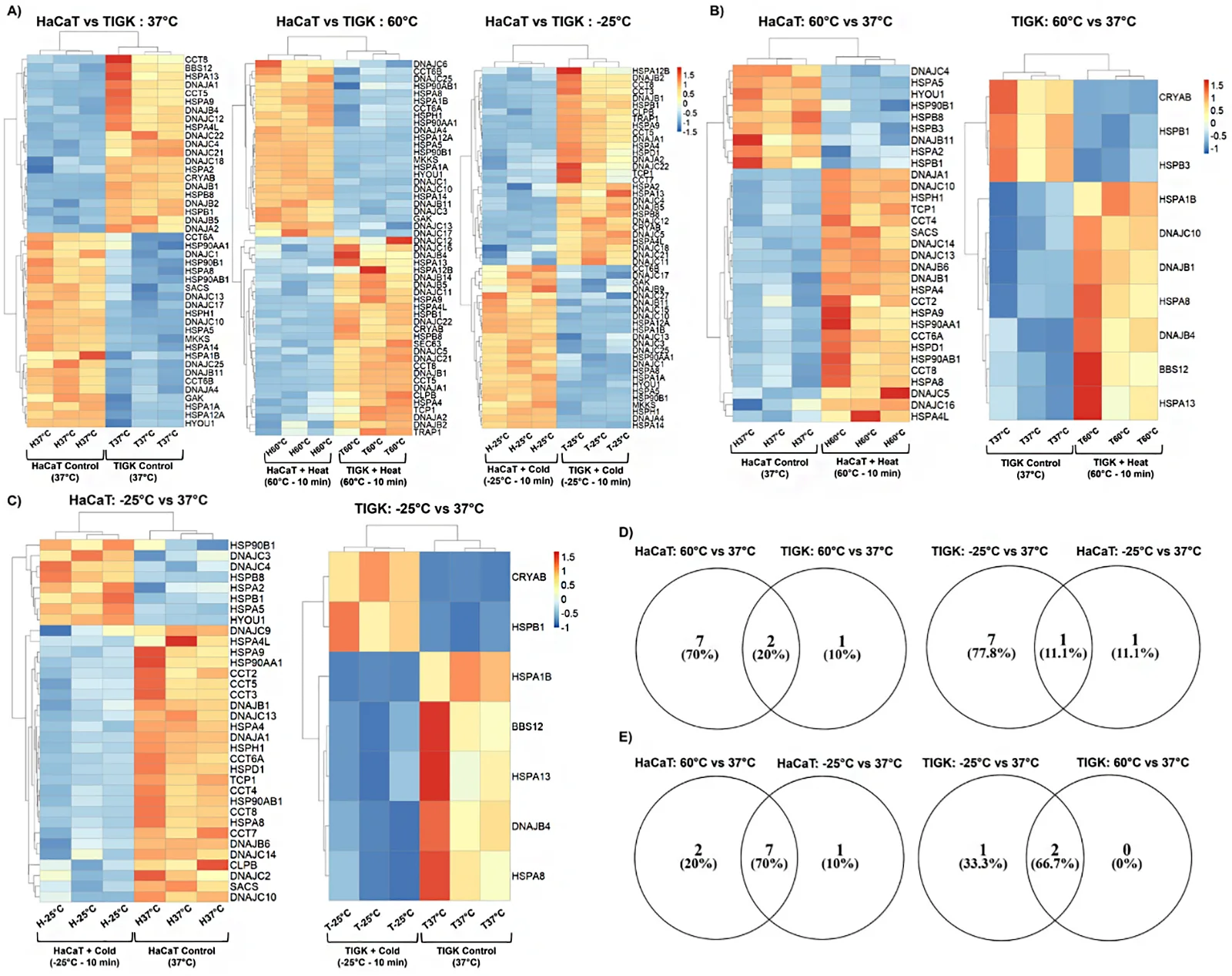
This study investigated if skin and oral keratinocytes have differential responses to heat- and cold-induced injury. Oral keratinocytes (TIGKs) were found to exhibit an enhanced viability following heat-induced injury compared to skin keratinocytes (HaCaTs). However, there were no discernible differences between skin and oral keratinocyte viability following cold-induced injury. To examine the transcriptomic differences between skin and oral keratinocytes in response to temperature-induced injury, an mRNA-sequencing gene expression dataset was generated. Differentially expressed genes (DEGs) including heat shock proteins (HSPs) were identified between HaCaTs and TIGKs at baseline (37 °C) and after heat- (60 °C) or cold-induced (−25 °C) injury. The comparative analyses suggest that skin and oral keratinocytes exhibit transcriptomic differences at baseline and in their responses to heat or cold exposure. The enhanced heat tolerance of TIGKs relative to HaCaTs may be due to an advantageous expression of a subset of HSPs at baseline in TIGKs.
These cells should be cultured in a specialized growth medium formulated for keratinocytes, supplemented with appropriate growth factors to promote optimal growth. Regular passaging and monitoring are also essential to maintain cell health and function. Detailed culture guidance is available for researchers.
Yes, Immortalized Human Gingival Keratinocytes-hTERT can be cryopreserved for long-term storage. It is recommended to follow established cryopreservation protocols to ensure high cell viability upon thawing.
Unlike primary gingival keratinocytes, which have a limited lifespan and can lose functionality over time, Immortalized Human Gingival Keratinocytes-hTERT maintain consistent growth and stability. This allows for repeated experiments and screening without the variability associated with primary cell cultures.
Ask a Question
Write your own review

Description: Nasal epithelial cells form the outermost protective layer against environmental factors. They clean, humidify, and warm inhaled air. They produces mucus, which binds particles that are subsequently ...
Description: Immortalized Human Splenic Endothelial Cells-SV40 have been obtained immortalizing Human Splenic Endothelial Cells with SV40LT expressing lentiviral particles. Immortalized cells were controlled ...
Description: Immortalized Human Corneal Epithelial Cells-SV40 have been obtained immortalizing Human Corneal Epithelial Cells with Lenti-SV40 Lentivirus. Immortalized cells were controlled passaging side by side ...
Description: Immortalized Human Lymphatic Endothelial Cells-SV40 were developed from human tissues transduced with a lentiviral expression vector containing the SV40T gene. The cell line was continuously cultured ...
- Adipose Tissue-Derived Stem Cells
- Human Neurons
- Mouse Probe
- Whole Chromosome Painting Probes
- Hepatic Cells
- Renal Cells
- In Vitro ADME Kits
- Tissue Microarray
- Tissue Blocks
- Tissue Sections
- FFPE Cell Pellet
- Probe
- Centromere Probes
- Telomere Probes
- Satellite Enumeration Probes
- Subtelomere Specific Probes
- Bacterial Probes
- ISH/FISH Probes
- Exosome Isolation Kit
- Human Adult Stem Cells
- Mouse Stem Cells
- iPSCs
- Mouse Embryonic Stem Cells
- iPSC Differentiation Kits
- Mesenchymal Stem Cells
- Immortalized Human Cells
- Immortalized Murine Cells
- Cell Immortalization Kit
- Adipose Cells
- Cardiac Cells
- Dermal Cells
- Epidermal Cells
- Peripheral Blood Mononuclear Cells
- Umbilical Cord Cells
- Monkey Primary Cells
- Mouse Primary Cells
- Breast Tumor Cells
- Colorectal Tumor Cells
- Esophageal Tumor Cells
- Lung Tumor Cells
- Leukemia/Lymphoma/Myeloma Cells
- Ovarian Tumor Cells
- Pancreatic Tumor Cells
- Mouse Tumor Cells