

Immortalized Human Gingival Fibroblasts-SV40
Cat.No.: CSC-I2120Z
Species: homo sapiens
Morphology: Polygonal
Culture Properties: Adherent
- Specification
- Background
- Scientific Data
- Q & A
- Customer Review
free from contaminations (bacteria incl. mycoplasma, fungi, HIV, HAV, HBV, HCV, Parvo-B19) and cross-contaminations
Note: Never can cells be kept at -20°C.
Immortalized Human Gingival Fibroblasts-SV40 are human gingival connective tissue cells immortalized by introduction of the SV40 Large T antigen, which confers extended proliferative capacity while retaining important biological characteristics of primary gingival fibroblasts. These cells have a typical spindle structure of fibroblasts and retain the ability to produce components of the extracellular matrix (ECM), respond to inflammatory stimuli, and participate in tissue remodeling processes.
Gingival fibroblasts are one of the principal cell types of gingival connective tissue and play a key role in the maintenance of homeostasis of the periodontal tissue, in the regulation of wound healing and in mediating host responses to microbial infection. The SV40 immortalization process allows to bypass the short life span of primordial cells and provides a stable and repeatable in vitro model for long-term investigations and high-throughput studies.
Immortalized Human Gingival Fibroblasts-SV40 are commonly employed in periodontal disease research, oral inflammatory studies, biomaterial evaluation and regenerative dentistry. They are useful instruments for studying host-pathogen interactions with oral bacteria, cytokine signaling pathways, extracellular matrix remodeling, and fibrosis-related mechanisms. These cells are also widely used to evaluate the biocompatibility of dental materials, implant surface changes and tissue engineering scaffolds.
In Vitro Cytotoxicity of Composite Resins on Human Gingival Fibroblasts
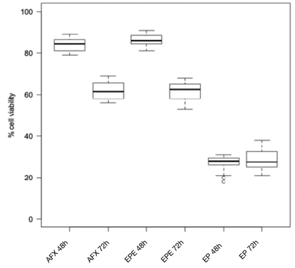
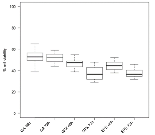
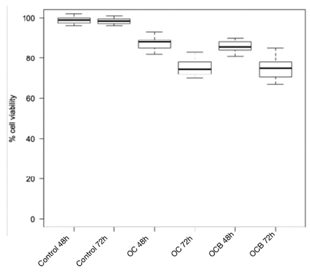
Beltrami et al. assessed the cytotoxicity of eight composite resins using immortalized human gingival fibroblasts (HGF-1). Intragroup analysis revealed that cell viability was significantly decreased after 72 h of incubation compared to 48 h for all resins except Enamel Plus HRi and G-aenial (Figs. 1,2). Enamel Plus HRi was highly toxic and G-aenial somewhat toxic at 48 h with no change in viability at 72 h.
The lowest cytotoxicity (viability > 80%) was seen for Omnichroma, Omnichroma Blocker, Admira Fusion x-tra, and Enamel Plus HRi Bio Function Enamel at 48 h, without significant differences between groups (Fig. 3). At 72 h, Omnichroma and Omnichroma Blocker still exhibited modest cytotoxicity, but the viability was dramatically decreased as compared with 48 h. Admira Fusion x-tra and Enamel Plus HRi Bio Function Enamel showed no to moderate cytotoxicity with no significant intergroup difference. G-aenial Flo X and Enamel Plus HRi Bio Function Bio Dentine revealed similar toxicity profiles at both time points, although with a lower survivability at 72 h than at 48 h.
Ask a Question
Write your own review
Description: Nasal epithelial cells form the outermost protective layer against environmental factors. They clean, humidify, and warm inhaled air. They produces mucus, which binds particles that are subsequently ...

Description: Immortalized Human Splenic Endothelial Cells-SV40 have been obtained immortalizing Human Splenic Endothelial Cells with SV40LT expressing lentiviral particles. Immortalized cells were controlled ...
Description: Immortalized Human Corneal Epithelial Cells-SV40 have been obtained immortalizing Human Corneal Epithelial Cells with Lenti-SV40 Lentivirus. Immortalized cells were controlled passaging side by side ...
Description: Immortalized Human Lymphatic Endothelial Cells-SV40 were developed from human tissues transduced with a lentiviral expression vector containing the SV40T gene. The cell line was continuously cultured ...
- Adipose Tissue-Derived Stem Cells
- Human Neurons
- Mouse Probe
- Whole Chromosome Painting Probes
- Hepatic Cells
- Renal Cells
- In Vitro ADME Kits
- Tissue Microarray
- Tissue Blocks
- Tissue Sections
- FFPE Cell Pellet
- Probe
- Centromere Probes
- Telomere Probes
- Satellite Enumeration Probes
- Subtelomere Specific Probes
- Bacterial Probes
- ISH/FISH Probes
- Exosome Isolation Kit
- Human Adult Stem Cells
- Mouse Stem Cells
- iPSCs
- Mouse Embryonic Stem Cells
- iPSC Differentiation Kits
- Mesenchymal Stem Cells
- Immortalized Human Cells
- Immortalized Murine Cells
- Cell Immortalization Kit
- Adipose Cells
- Cardiac Cells
- Dermal Cells
- Epidermal Cells
- Peripheral Blood Mononuclear Cells
- Umbilical Cord Cells
- Monkey Primary Cells
- Mouse Primary Cells
- Breast Tumor Cells
- Colorectal Tumor Cells
- Esophageal Tumor Cells
- Lung Tumor Cells
- Leukemia/Lymphoma/Myeloma Cells
- Ovarian Tumor Cells
- Pancreatic Tumor Cells
- Mouse Tumor Cells