
Immortalized Human Foreskin Keratinocytes (16E6/E7 HFK)
Cat.No.: CSC-I9336L
Species: Homo sapiens
Source: Foreskin tissue
Morphology: Polygonal
Culture Properties: Adherent
- Specification
- Background
- Scientific Data
- Q & A
- Customer Review
Note: Never can cells be kept at -20 °C.
CIK-HT013 HT® Lenti-hTERT Immortalization Kit
CIK-HT003 HT® Lenti-SV40T Immortalization Kit
2) Microarray analysis of gene regulation
Immortalized Human Foreskin Keratinocytes (16E6/E7 HFK) are immortalized primary human foreskin keratinocytes transformed with human papillomavirus type 16 (HPV16) E6 and E7 oncogenes. Cultured 16E6/E7 HFK cells display the characteristic cobblestone morphology of stratified epithelial cells, growing in an adherent manner. Similar to primary human keratinocytes, they express keratinocyte markers including keratin 5 (KRT5), keratin 14 (KRT14), involucrin, and E-cadherin and are capable of partial differentiation. Additionally, they maintain some normal epithelial organization and responsiveness to differentiation factors.
16E6/E7 HFK cells have been used to study various aspects of HPV-mediated carcinogenesis, epithelial cell cycle regulation, and skin biology. In particular, they are used to study HPV oncogene function, DNA damage response, and transformation. These cells have also been used in studies involving barrier formation, inflammation, and drug responses.
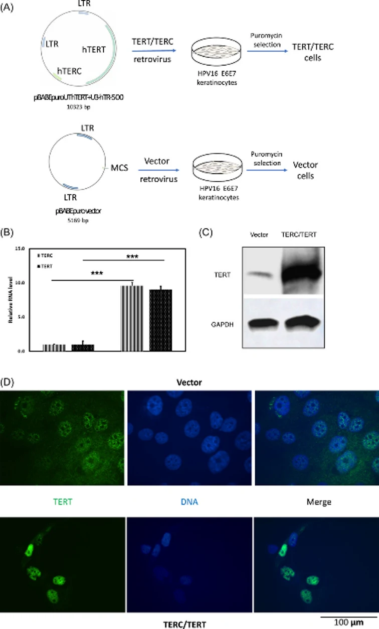
The Overexpression of hTERT/hTERC Increased Cell Growth Rate
Cervical cancer, the most common malignancy of the female genital tract, is associated with persistent high-risk HPV infection. The viral oncoproteins E6 and E7 cooperatively immortalize cervical cells but are insufficient for full tumorigenicity. During progression from dysplasia to carcinoma, telomerase components TERT and TERC are activated and amplified. Wang's team investigated whether elevated telomerase mediates acquisition of the tumorigenic phenotype.
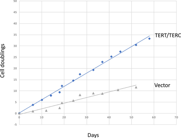
Immortalized human foreskin keratinocytes transduced with HPV16 E6/E7 (HFK/E6E7) were infected with control vector or retrovirus co-expressing hTERT/hTERC (Fig. 1A). Puromycin-selected cells were designated HFK/E6E7+vector and HFK/E6E7+hTERT/hTERC. Overexpression was confirmed at RNA (Fig. 1B) and protein levels (Fig. 1C), with nuclear hTERT localization by immunofluorescence (Fig. 1D). HFK/E6E7+hTERT/hTERC cells exhibited significantly faster growth (1.36 days/doubling) compared to controls (3.40 days/doubling) (Fig. 1E).
Ask a Question
Write your own review

Description: Nasal epithelial cells form the outermost protective layer against environmental factors. They clean, humidify, and warm inhaled air. They produces mucus, which binds particles that are subsequently ...

Description: Immortalized Human Splenic Endothelial Cells-SV40 have been obtained immortalizing Human Splenic Endothelial Cells with SV40LT expressing lentiviral particles. Immortalized cells were controlled ...
Description: Immortalized Human Corneal Epithelial Cells-SV40 have been obtained immortalizing Human Corneal Epithelial Cells with Lenti-SV40 Lentivirus. Immortalized cells were controlled passaging side by side ...
Description: Immortalized Human Lymphatic Endothelial Cells-SV40 were developed from human tissues transduced with a lentiviral expression vector containing the SV40T gene. The cell line was continuously cultured ...
- Adipose Tissue-Derived Stem Cells
- Human Neurons
- Mouse Probe
- Whole Chromosome Painting Probes
- Hepatic Cells
- Renal Cells
- In Vitro ADME Kits
- Tissue Microarray
- Tissue Blocks
- Tissue Sections
- FFPE Cell Pellet
- Probe
- Centromere Probes
- Telomere Probes
- Satellite Enumeration Probes
- Subtelomere Specific Probes
- Bacterial Probes
- ISH/FISH Probes
- Exosome Isolation Kit
- Human Adult Stem Cells
- Mouse Stem Cells
- iPSCs
- Mouse Embryonic Stem Cells
- iPSC Differentiation Kits
- Mesenchymal Stem Cells
- Immortalized Human Cells
- Immortalized Murine Cells
- Cell Immortalization Kit
- Adipose Cells
- Cardiac Cells
- Dermal Cells
- Epidermal Cells
- Peripheral Blood Mononuclear Cells
- Umbilical Cord Cells
- Monkey Primary Cells
- Mouse Primary Cells
- Breast Tumor Cells
- Colorectal Tumor Cells
- Esophageal Tumor Cells
- Lung Tumor Cells
- Leukemia/Lymphoma/Myeloma Cells
- Ovarian Tumor Cells
- Pancreatic Tumor Cells
- Mouse Tumor Cells