

Immortalized Human Esophageal Epithelial Cells-SV40 (Het-1A)
Cat.No.: CSC-I2303Z
Morphology: Polygonal
Culture Properties: Adherent
- Specification
- Background
- Scientific Data
- Q & A
- Customer Review
Immortalized human esophageal epithelial cells-SV40 (Het-1A) are established by transfecting normal human esophageal autopsy tissue with plasmid pRSV-T, which contains the Rous sarcoma virus long terminal repeat (RSV-LTR) promoter driving the simian virus 40 (SV40) large T-antigen gene. The advantages of Het-1A cells are fourfold. First, extended proliferative capacity – following a crisis period of 6–8 months, Het-1A emerged as a continuously growing line that has undergone more than 250 population doublings, with a doubling time of approximately 27.4 hours. This addresses the fundamental supply constraints of primary cultures. Second, retention of epithelial characteristics – Het-1A cells maintain epithelial morphology, stain positively for cytokeratins, and express the SV40 T-antigen. They remain responsive to physiological regulators: proliferation is stimulated by Ca2+ and inhibited by fetal bovine serum, transforming growth factor-beta 1 and 2. Third, non-tumorigenicity – Het-1A cells have remained non-tumorigenic in athymic nude mice for more than 12 months, confirming they are immortalized but not transformed, making them an appropriate normal control for carcinogenesis studies. Fourth, experimental reproducibility and scalability – the immortalized nature eliminates donor-to-donor variability inherent to primary cultures, enabling consistent, repeatable experiments and high-throughput screening. Het-1A cells are extensively used to model esophageal diseases including Barrett's esophagus, gastroesophageal reflux disease (GERD), and esophageal cancer, as well as to investigate responses to acid and bile reflux, oxidative stress, and chemopreventive agents.
The Effect of Thymic Stromal Lymphopoietin on Human Squamous Esophageal Cell Line HET-1A
Reflux esophagitis (RE) is characterized by the infiltration of inflammatory cells into the damaged squamous epithelium. Thymic stromal lymphopoietin (TSLP) has been implicated in promoting T helper type 2 (Th2) inflammation. This study investigates the protective role of TSLP downregulation in RE.
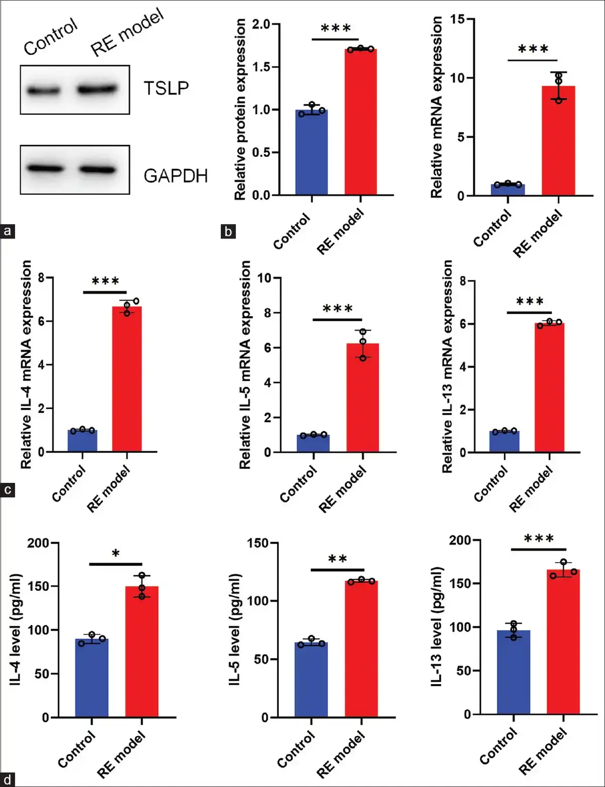
HET-1A cells were exposed to an acid and bile mixture to construct a model of RE. Then, the western blot showed that exposure to acid and bile mixture significantly increased the expression of TSLP in HET-1A cells [Fig. 1a]. qPCR results were consistent with western blot results [Fig. 1b]. Moreover, exposure to the acid–bile mixture significantly increased the expression levels of IL-4, IL-5, and IL-13 in HET-1A cells and supernatant [Fig. 1d]. The above results indicate that TSLP expression was increased in the RE cell model and that TSLP is involved in RE.
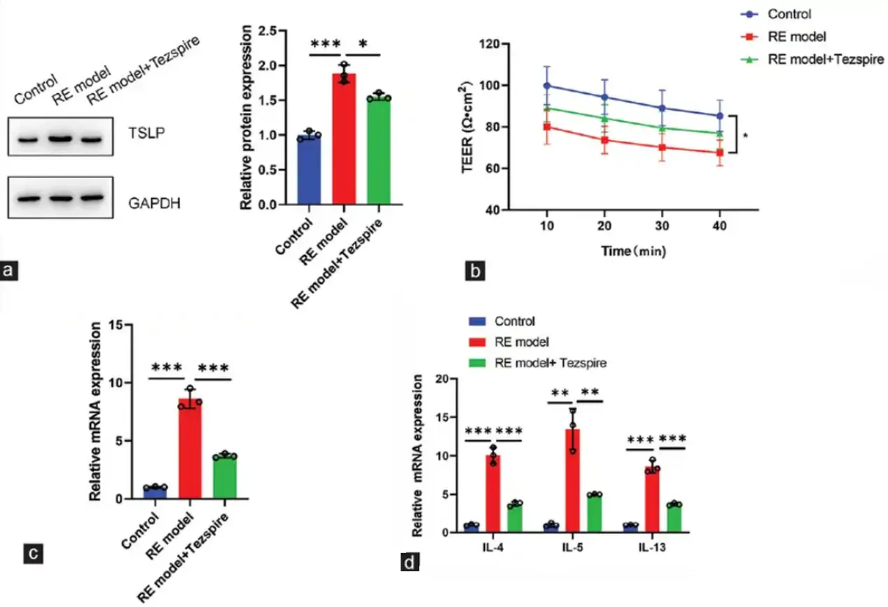
Tezspire demonstrates a high affinity for human TSLP and exerts inhibitory effects on multiple downstream inflammatory pathways. To evaluate the potential of targeting TSLP as a therapeutic strategy for RE, we administered Tezspire to HET-1A cells and conducted a series of analyses, including western blot, qPCR, TEER measurements, and ELISA. The findings revealed that Tezspire significantly reduced TSLP expression [Fig. 2a and c]. In addition, Tezspire markedly enhanced the barrier tightness of HET-1A cells subjected to an acid and bile mixture [Fig. 2b]. Furthermore, Tezspire effectively diminished the expression of pro-inflammatory factors, as demonstrated in Fig. 2d.
Ask a Question
Write your own review
Description: Nasal epithelial cells form the outermost protective layer against environmental factors. They clean, humidify, and warm inhaled air. They produces mucus, which binds particles that are subsequently ...
Description: Immortalized Human Splenic Endothelial Cells-SV40 have been obtained immortalizing Human Splenic Endothelial Cells with SV40LT expressing lentiviral particles. Immortalized cells were controlled ...
Description: Immortalized Human Corneal Epithelial Cells-SV40 have been obtained immortalizing Human Corneal Epithelial Cells with Lenti-SV40 Lentivirus. Immortalized cells were controlled passaging side by side ...

Description: Immortalized Human Lymphatic Endothelial Cells-SV40 were developed from human tissues transduced with a lentiviral expression vector containing the SV40T gene. The cell line was continuously cultured ...
- Adipose Tissue-Derived Stem Cells
- Human Neurons
- Mouse Probe
- Whole Chromosome Painting Probes
- Hepatic Cells
- Renal Cells
- In Vitro ADME Kits
- Tissue Microarray
- Tissue Blocks
- Tissue Sections
- FFPE Cell Pellet
- Probe
- Centromere Probes
- Telomere Probes
- Satellite Enumeration Probes
- Subtelomere Specific Probes
- Bacterial Probes
- ISH/FISH Probes
- Exosome Isolation Kit
- Human Adult Stem Cells
- Mouse Stem Cells
- iPSCs
- Mouse Embryonic Stem Cells
- iPSC Differentiation Kits
- Mesenchymal Stem Cells
- Immortalized Human Cells
- Immortalized Murine Cells
- Cell Immortalization Kit
- Adipose Cells
- Cardiac Cells
- Dermal Cells
- Epidermal Cells
- Peripheral Blood Mononuclear Cells
- Umbilical Cord Cells
- Monkey Primary Cells
- Mouse Primary Cells
- Breast Tumor Cells
- Colorectal Tumor Cells
- Esophageal Tumor Cells
- Lung Tumor Cells
- Leukemia/Lymphoma/Myeloma Cells
- Ovarian Tumor Cells
- Pancreatic Tumor Cells
- Mouse Tumor Cells