

Immortalized Human Endometrial Stromal Cells-SV40
Cat.No.: CSC-I9354L
Species: Homo sapiens
Source: Reproductive system
Morphology: Polygonal
Culture Properties: Adherent
- Specification
- Background
- Scientific Data
- Q & A
- Customer Review
Note: Never can cells be kept at -20 °C.
The human endometrium undergoes dynamic cyclic remodeling, and its stromal compartment plays essential roles in menstruation, decidualization, and embryo implantation. Obtaining primary human endometrial stromal cells (hESCs) is challenging due to tissue scarcity and their limited proliferative capacity in culture, which constrains long-term investigations. To overcome these limitations, human endometrial stromal cells have been immortalized via stable introduction of the Simian Virus 40 (SV40) large T antigen.
SV40 large T antigen disrupts p53- and pRB-dependent cell cycle checkpoints, enabling the cells to bypass replicative senescence and achieve continuous proliferation. The resulting immortalized hESC line retains key morphological characteristics of primary stromal cells, exhibiting an adherent, polygonal morphology. Importantly, these cells maintain expression of stromal lineage markers-including vimentin, smooth muscle actin, and desmin-as confirmed by immunostaining and Western blot analysis, demonstrating that immortality is achieved without loss of the original phenotype. Furthermore, immortalized cells preserve functional responsiveness to physiological stimuli, such as hormonal induction of decidualization, thereby recapitulating core biological behaviors of their primary counterparts.
Collectively, the Immortalized Human Endometrial Stromal Cells-SV40 line provides a consistent, reproducible, and phenotypically faithful model system for studying endometrial physiology, hormonal regulation, and the pathogenesis of endometriosis and endometrial cancer, while circumventing the material limitations and passage constraints inherent to primary cell cultures.
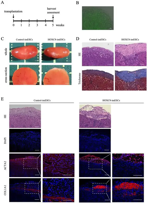
Establishment of A Xenograft Model of Endometriosis-Associated Fibrosis Using Human Immortalized Endometrial Stromal Cells Overexpressing HOXC8
Endometriosis is a disease characterized by fibrosis and adhesions. There are still no treatment methods targeting these conditions. One reason for this is the lack of useful animal models to investigate the mechanisms of fibrosis and adhesion in endometriosis. Takasaki-Kawasaki, Hitomi, et al. previously proposed that Homeobox C8 (HOXC8) acts as an upstream regulator involved in endometriosis development, promoting fibrosis by activating transforming growth factor β (TGFB) signaling in vitro. This study was undertaken to establish a xenograft mouse model for endometriosis-associated fibrosis using HOXC8-overexpressing cells.
Stable lines of immortalized human endometrial stromal cells overexpressing HOXC8 (HOXC8-imESCs) and mock control lines (Control-imESCs) were established and evaluated for cellular functions in vitro. These cell lines were then transplanted under the renal capsule of severely immunodeficient mice, and the xenografts were harvested five weeks after transplantation. In vitro, cell migration, invasion, and fibrotic capacity were enhanced in HOXC8-imESCs compared to Control-imESCs. In the xenografts, HOXC8-imESCs exhibited significant collagen fiber production compared to Control-imESCs. Additionally, immunohistochemistry of the xenografts detected phosphorylated SMAD2/3 exclusively in HOXC8-imESCs, indicating that HOXC8 overexpression activates the TGFB/SMAD pathway aberrantly in vivo.
Ask a Question
Write your own review
Description: Nasal epithelial cells form the outermost protective layer against environmental factors. They clean, humidify, and warm inhaled air. They produces mucus, which binds particles that are subsequently ...
Description: Immortalized Human Splenic Endothelial Cells-SV40 have been obtained immortalizing Human Splenic Endothelial Cells with SV40LT expressing lentiviral particles. Immortalized cells were controlled ...

Description: Immortalized Human Corneal Epithelial Cells-SV40 have been obtained immortalizing Human Corneal Epithelial Cells with Lenti-SV40 Lentivirus. Immortalized cells were controlled passaging side by side ...
Description: Immortalized Human Lymphatic Endothelial Cells-SV40 were developed from human tissues transduced with a lentiviral expression vector containing the SV40T gene. The cell line was continuously cultured ...
- Adipose Tissue-Derived Stem Cells
- Human Neurons
- Mouse Probe
- Whole Chromosome Painting Probes
- Hepatic Cells
- Renal Cells
- In Vitro ADME Kits
- Tissue Microarray
- Tissue Blocks
- Tissue Sections
- FFPE Cell Pellet
- Probe
- Centromere Probes
- Telomere Probes
- Satellite Enumeration Probes
- Subtelomere Specific Probes
- Bacterial Probes
- ISH/FISH Probes
- Exosome Isolation Kit
- Human Adult Stem Cells
- Mouse Stem Cells
- iPSCs
- Mouse Embryonic Stem Cells
- iPSC Differentiation Kits
- Mesenchymal Stem Cells
- Immortalized Human Cells
- Immortalized Murine Cells
- Cell Immortalization Kit
- Adipose Cells
- Cardiac Cells
- Dermal Cells
- Epidermal Cells
- Peripheral Blood Mononuclear Cells
- Umbilical Cord Cells
- Monkey Primary Cells
- Mouse Primary Cells
- Breast Tumor Cells
- Colorectal Tumor Cells
- Esophageal Tumor Cells
- Lung Tumor Cells
- Leukemia/Lymphoma/Myeloma Cells
- Ovarian Tumor Cells
- Pancreatic Tumor Cells
- Mouse Tumor Cells