

Immortalized Human Chondrocytes-hTERT
Cat.No.: CSC-I9042L
Species: Homo sapiens
Source: Normal human articular cartilage
Morphology: Multipolar
Culture Properties: Adherent
- Specification
- Background
- Scientific Data
- Q & A
- Customer Review
Note: Never can cells be kept at -20 °C.
CIK-HT013 HT® Lenti-hTERT Immortalization Kit
An immortalized human chondrocyte line was generated via stable overexpression of human telomerase reverse transcriptase (hTERT). Unlike SV40T-immortalized cells, hTERT-mediated immortalization does not disrupt the p53 or pRB pathways, thereby maintaining genomic integrity and normal DNA damage responses while enabling extended replicative lifespan without oncogenic transformation.
These immortalized hTERT-chondrocytes possess the following advantages: (1) preservation of the chondrocyte phenotype, including typical polygonal morphology and expression of SOX9, COL2A1, and aggrecan, as confirmed by RT-PCR, Western blot, and immunostaining; (2) retention of ECM-forming capacity in both monolayer and three-dimensional culture systems; (3) normal diploid karyotype without chromosomal aberrations; (4) lack of tumorigenicity in immunodeficient mice; (5) functional responsiveness to pro-inflammatory cytokines (IL-1β, TNF-α) and anabolic/catabolic stimuli; and (6) stable behavior for >50 population doublings.
Collectively, Immortalized Human Chondrocytes-hTERT provide an unlimited, phenotypically stable, and reproducible in vitro model for chondrocyte biology, osteoarthritis drug discovery, and cartilage tissue engineering, circumventing the dedifferentiation and scarcity constraints of primary chondrocyte cultures.
Generation of Human Immortalized Chondrocytes from Osteoarthritic and Healthy Cartilage
After a few passages of in vitro culture, primary human articular chondrocytes undergo senescence and loss of their phenotype. Most of the available chondrocyte cell lines have been obtained from cartilage tissues different from diarthrodial joints, and their utility for osteoarthritis (OA) research is reduced. This study aims to establish immortalized chondrocyte cell lines proceeded from the articular cartilage of patients with and without OA.
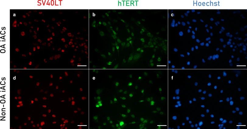
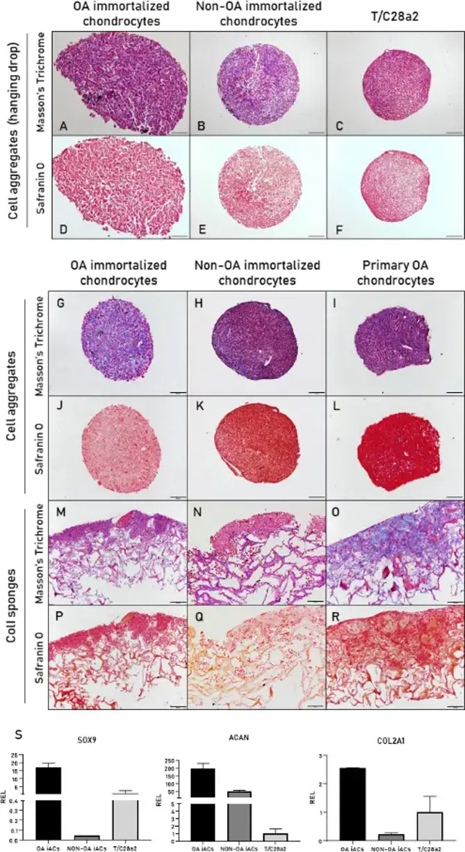
Primary articular chondrocytes were transduced with telomerase reverse transcriptase (hTERT) and SV40 large T antigen (SV40LT). Proliferative capacity, degree of senescence, and chondrocyte surface antigen expression in transduced chondrocytes were evaluated. Furthermore, their capacity to synthesize a tissue similar to cartilage and to respond to interleukin (IL)-1β was assessed.
Co-expression of both transgenes (SV40 and hTERT) were observed in the nuclei of transduced chondrocytes. Generated chondrocyte cell lines showed a high proliferation capacity and less than 2% of senescent cells. These cell lines were able to form 3D aggregates analogous to those generated by primary articular chondrocytes, but were unsuccessful in synthesizing cartilage-like tissue when seeded on type I collagen sponges. However, generated chondrocyte cell lines maintained the potential to respond to IL-1β stimulation.
Ask a Question
Write your own review

Description: Nasal epithelial cells form the outermost protective layer against environmental factors. They clean, humidify, and warm inhaled air. They produces mucus, which binds particles that are subsequently ...
Description: Immortalized Human Splenic Endothelial Cells-SV40 have been obtained immortalizing Human Splenic Endothelial Cells with SV40LT expressing lentiviral particles. Immortalized cells were controlled ...
Description: Immortalized Human Corneal Epithelial Cells-SV40 have been obtained immortalizing Human Corneal Epithelial Cells with Lenti-SV40 Lentivirus. Immortalized cells were controlled passaging side by side ...
Description: Immortalized Human Lymphatic Endothelial Cells-SV40 were developed from human tissues transduced with a lentiviral expression vector containing the SV40T gene. The cell line was continuously cultured ...
- Adipose Tissue-Derived Stem Cells
- Human Neurons
- Mouse Probe
- Whole Chromosome Painting Probes
- Hepatic Cells
- Renal Cells
- In Vitro ADME Kits
- Tissue Microarray
- Tissue Blocks
- Tissue Sections
- FFPE Cell Pellet
- Probe
- Centromere Probes
- Telomere Probes
- Satellite Enumeration Probes
- Subtelomere Specific Probes
- Bacterial Probes
- ISH/FISH Probes
- Exosome Isolation Kit
- Human Adult Stem Cells
- Mouse Stem Cells
- iPSCs
- Mouse Embryonic Stem Cells
- iPSC Differentiation Kits
- Mesenchymal Stem Cells
- Immortalized Human Cells
- Immortalized Murine Cells
- Cell Immortalization Kit
- Adipose Cells
- Cardiac Cells
- Dermal Cells
- Epidermal Cells
- Peripheral Blood Mononuclear Cells
- Umbilical Cord Cells
- Monkey Primary Cells
- Mouse Primary Cells
- Breast Tumor Cells
- Colorectal Tumor Cells
- Esophageal Tumor Cells
- Lung Tumor Cells
- Leukemia/Lymphoma/Myeloma Cells
- Ovarian Tumor Cells
- Pancreatic Tumor Cells
- Mouse Tumor Cells