

Immortalized Human Aortic Endothelial Cells
Cat.No.: CSC-I2053Z
Species: homo sapiens
Morphology: Polygonal
Culture Properties: Adherent
- Specification
- Background
- Scientific Data
- Q & A
- Customer Review
free from contaminations (bacteria incl. mycoplasma, fungi, HIV, HAV, HBV, HCV, Parvo-B19) and cross-contaminations
Note: Never can cells be kept at -20°C.
Immortalized human aortic endothelial cells (HAECs) are established by transducing primary HAECs with recombinant lentiviruses carrying the simian virus 40 (SV40) large T antigen or by stably expressing human telomerase reverse transcriptase (hTERT). The advantages of immortalized HAECs are fourfold. First, extended proliferative capacity – whereas primary cells senesce within a few passages, SV40-transduced cells can be cultured beyond 30 passages, with a population doubling time of approximately 34–44 hours, providing a consistent and virtually unlimited cell supply. Second, phenotypic fidelity – immortalized HAECs retain key endothelial characteristics, including cobblestone morphology, expression of endothelial markers such as CD31/PECAM-1, von Willebrand factor (vWF), and VE-cadherin, as well as functional attributes including LDL uptake and formation of tubular structures that mimic natural blood vessel development. They respond to physiological stimuli: proliferation increases upon VEGF stimulation, and treatment with TNFα induces an effective inflammatory response with upregulation of adhesion molecules ICAM-1 and VCAM-1. Third, functional relevance – these cells produce both antithrombotic and thrombotic factors such as tissue plasminogen activator (t-PA) and plasminogen activator inhibitor-1 (PAI-1), and maintain nitric oxide (NO) production. hTERT-immortalized lines exhibit greater eNOS expression and NO activity compared to senescent primary cells. When co-cultured with fibroblasts, they can form neoangiogenic tubular networks responsive to VEGF stimulation and suramin inhibition. Fourth, experimental reproducibility and scalability – the immortalized nature eliminates donor-to-donor variability inherent to primary cultures, enabling consistent, repeatable experiments and high-throughput screening for drug development and cardiovascular disease research. They are amenable to genetic modification including CRISPR-based editing, and have been extensively validated in studies of atherosclerosis, hypertension, thrombosis, angiogenesis, and stent graft compatibility.
PCSK9 Inhibition Ameliorates Microplastic-Induced Endothelial Redox Imbalance via SIRT6 Modulation
This study aims to investigate the effects of microplastics (MPs) on endothelial cell function and redox state and the underlying mechanisms. Immortalized human aortic endothelial cells (teloHAEC) were treated with MPs in the form of polyethylene (PE) and polyvinyl chloride (PVC) alone or combined PE + PVC (PE + PVC) for up to 48 h.
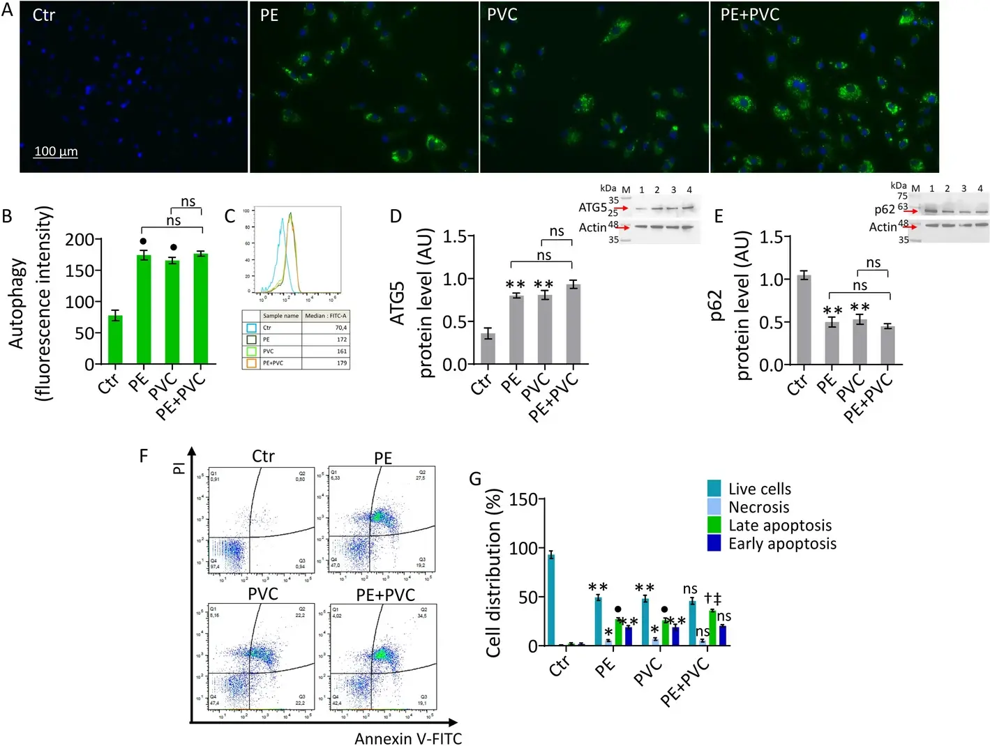
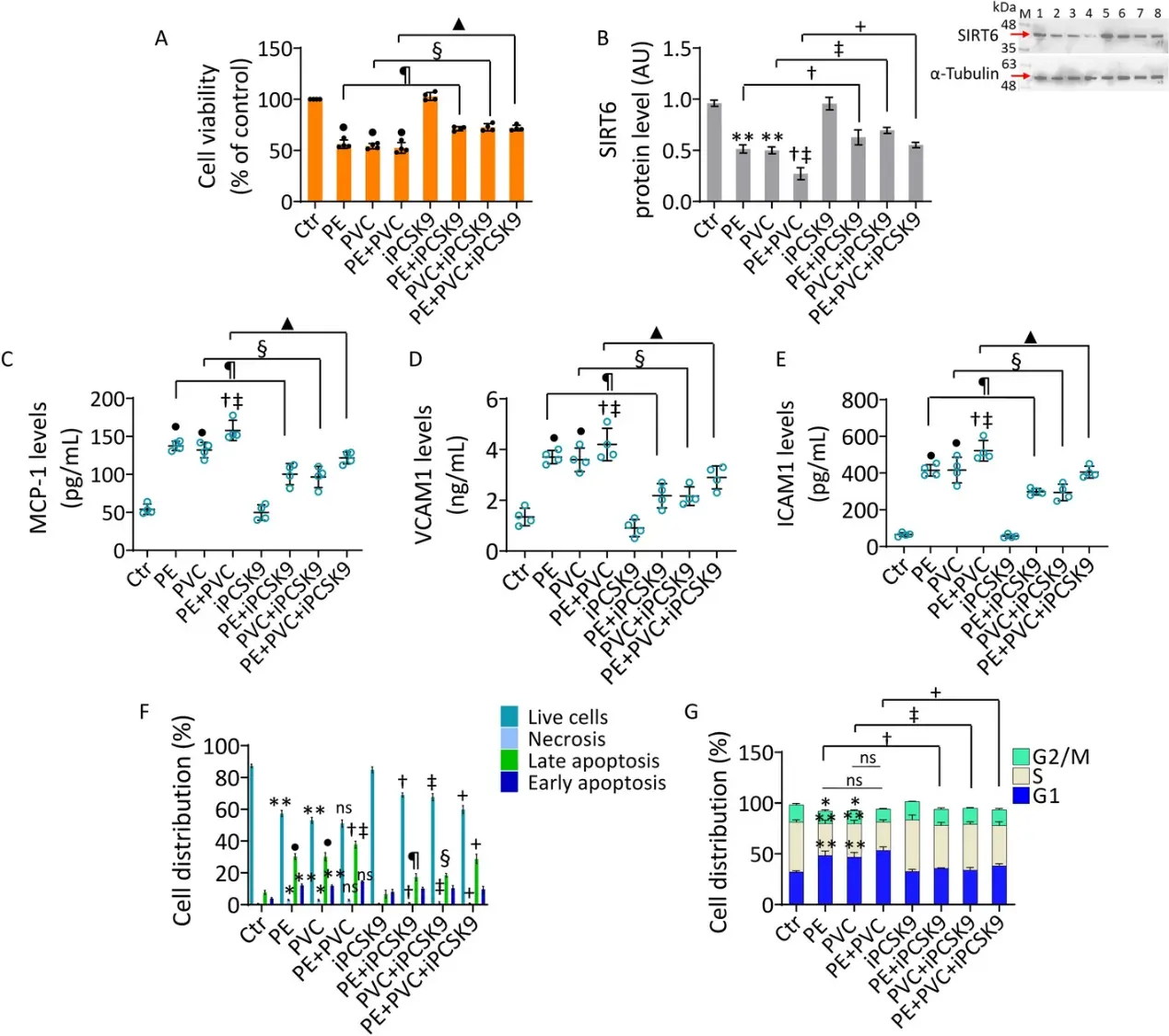
The results demonstrated that PE and PVC, alone or in combination, upregulated inflammatory mediators monocyte chemoattractant protein-1 (MCP-1), vascular cell adhesion molecule-1 (VCAM1), and intercellular adhesion molecule-1 (ICAM1), modulated the expression of autophagy markers anti-autophagy related 5 (ATG5) and p62, impaired mitochondrial metabolism by reducing maximal and basal respiration and adenosine triphosphate (ATP) production, promoted reactive oxygen species (ROS) accumulation and cell cycle perturbations, and increased apoptosis cell death. These events were accompanied by a downregulation of sirtuin 6 (SIRT6) expression and an upregulation of PCSK9, at protein and messenger RNA (mRNA) levels.
Treatment with the PCSK9 inhibitor (iPCSK9) evolocumab ameliorated MP-induced cellular redox state imbalance, mitochondrial metabolism alteration, and SIRT6 downregulated levels. SIRT6 transient silencing experiments denied the beneficial effects of iPCSK9 treatment, indicating that the pleiotropic functions of iPCSK9 may occur, at least in part, via modulation of SIRT6 and Forkhead box O3 (FOXO3A) expression levels.
Ask a Question
Write your own review

Description: Nasal epithelial cells form the outermost protective layer against environmental factors. They clean, humidify, and warm inhaled air. They produces mucus, which binds particles that are subsequently ...
Description: Immortalized Human Splenic Endothelial Cells-SV40 have been obtained immortalizing Human Splenic Endothelial Cells with SV40LT expressing lentiviral particles. Immortalized cells were controlled ...
Description: Immortalized Human Corneal Epithelial Cells-SV40 have been obtained immortalizing Human Corneal Epithelial Cells with Lenti-SV40 Lentivirus. Immortalized cells were controlled passaging side by side ...
Description: Immortalized Human Lymphatic Endothelial Cells-SV40 were developed from human tissues transduced with a lentiviral expression vector containing the SV40T gene. The cell line was continuously cultured ...
- Adipose Tissue-Derived Stem Cells
- Human Neurons
- Mouse Probe
- Whole Chromosome Painting Probes
- Hepatic Cells
- Renal Cells
- In Vitro ADME Kits
- Tissue Microarray
- Tissue Blocks
- Tissue Sections
- FFPE Cell Pellet
- Probe
- Centromere Probes
- Telomere Probes
- Satellite Enumeration Probes
- Subtelomere Specific Probes
- Bacterial Probes
- ISH/FISH Probes
- Exosome Isolation Kit
- Human Adult Stem Cells
- Mouse Stem Cells
- iPSCs
- Mouse Embryonic Stem Cells
- iPSC Differentiation Kits
- Mesenchymal Stem Cells
- Immortalized Human Cells
- Immortalized Murine Cells
- Cell Immortalization Kit
- Adipose Cells
- Cardiac Cells
- Dermal Cells
- Epidermal Cells
- Peripheral Blood Mononuclear Cells
- Umbilical Cord Cells
- Monkey Primary Cells
- Mouse Primary Cells
- Breast Tumor Cells
- Colorectal Tumor Cells
- Esophageal Tumor Cells
- Lung Tumor Cells
- Leukemia/Lymphoma/Myeloma Cells
- Ovarian Tumor Cells
- Pancreatic Tumor Cells
- Mouse Tumor Cells