Hamster Hepatocytes
Cat.No.: CSC-C4760L
Species: Hamster
Source: Liver
Cell Type: Hepatocyte
- Specification
- Background
- Scientific Data
- Q & A
- Customer Review
Never can cryopreserved cells be kept at -22 °C.
Hamster hepatocytes are primary liver epithelial cells, and offer an in vitro hepatic model system to study metabolism, toxicology, and lipid biology. Isolated from the parenchymal compartment of the hamster liver, hepatocytes maintain many of the defining physiological attributes of mammalian hepatocytes, including a polygonal shape, large nuclei that are often binucleated, and lipid and glycogen storage in the cytoplasm. In cell culture, hepatocytes attach well to collagen-coated surfaces and retain metabolic activity for multiple days, including active Phase I and Phase II biotransformation mechanisms (CYP450-, UGT-, and SULT-mediated activities).
Hamster hepatocytes are also well suited to study of lipid and cholesterol metabolism. As hamsters are physiologically similar to humans in cholesterol metabolism, they can be used to study dyslipidemia, NAFLD, and lipid droplet formation. They also maintain glucose homeostasis, are responsive to insulin, and express a wide range of hepatic transporters. Hamster hepatocytes have been used for a number of applications in DMPK and hepatotoxicity, oxidative stress, and cross-species metabolic comparisons. They have also been used in studies of viral infection, broadening their applicability in the study of infectious diseases and antiviral drug discovery.
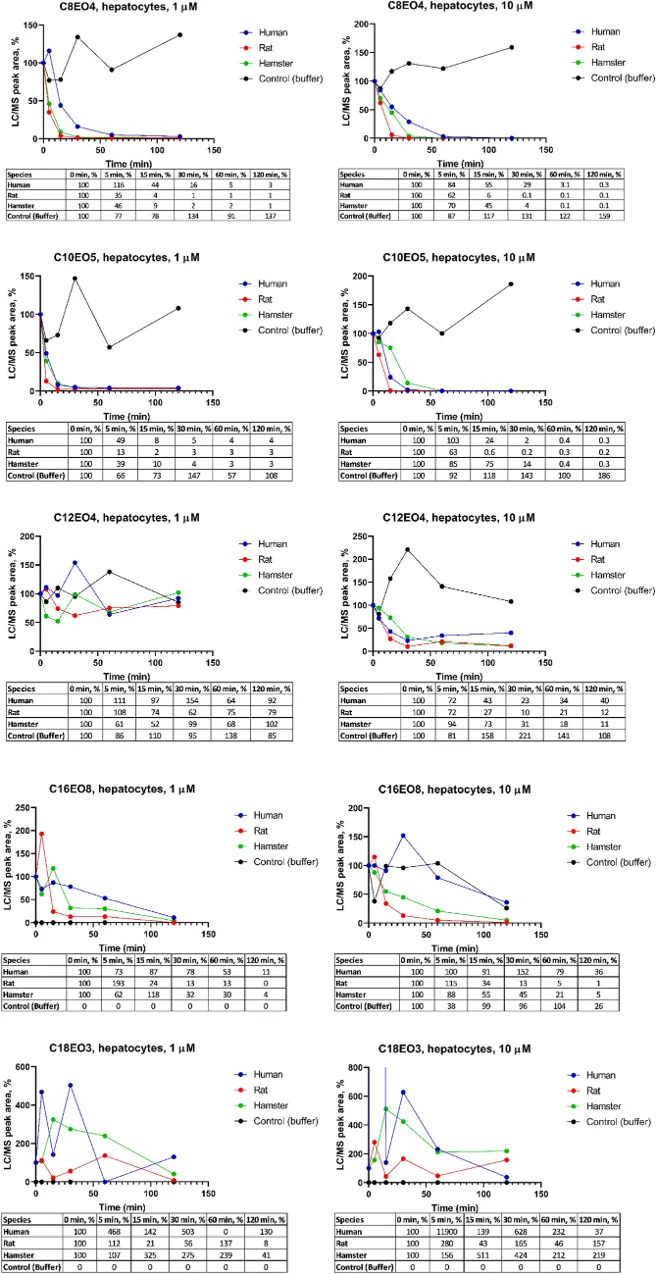
Metabolic Stability of Alcohol Ethoxylates (AEs) In Rat, Hamster, and Human Hepatocytes
Alcohol ethoxylates (AEs) are popular non-ionic surfactants. Here, Shi's team incubated five homologue AEs (C8EO4, C10EO5, C12EO4, C16EO8 and C18EO3) with human, rat, and hamster liver S9 fraction and hepatocytes to evaluate their in vitro metabolism.
The results of incubation with human, rat, and hamster hepatocytes are shown in Figure 1. Without cells, no disappearance was observed, but high variation in peak areas was seen with all compounds. In buffer incubations, C16EO8 at 1 μM and C18EO3 at both concentrations were not detected. For C8EO4 at 1 and 10 µM, only 0.1-3% remained after 120 min with hepatocytes. For C10EO5 at 1 and 10 µM, 0.2-4% remained. C12EO4 at 1 μM showed high fluctuation in LC/MS peak areas, but data at 10 µM were of good quality. After 120 min, 40% remained in human, 12% in rat, and 11% in hamster hepatocytes. For C16EO8 at 1 and 10 µM, 11-36% remained in human, 1-16% in rat, and 4-5% in hamster hepatocytes. C18EO3 showed high fluctuation in LC/MS peak areas, hindering further investigation.
Ask a Question
Write your own review
- You May Also Need
Description: Hamster Ovarian Smooth Muscle Cells are isolated from ovarian tissue of pathogen-free laboratory mice.
Description: Hamster Esophageal Epithelial Cells from Creative Bioarray are isolated from esophageal tissue of pathogen-free laboratory mice. Hamster Esophageal Epithelial Cells are grown in a T25 tissue culture ...
Description: Hamster Spleen Epithelial Cells from Creative Bioarray are isolated from spleen tissue of pathogen-free laboratory mice. Hamster Spleen Epithelial Cells are grown in a T25 tissue culture flask ...
Description: Hamster Corneal Epithelial Cells from Creative Bioarray are isolated from corneal tissue of pathogen-free laboratory mice. Hamster Corneal Epithelial Cells are grown in a T25 tissue culture flask ...
Description: Hamster Aortic Smooth Muscle Cells are isolated from aorta of hamster.
Description: Hamster Primary Pancreatic Fibroblasts are isolated from pancreas tissue of hamster.