Rat Pulmonary Artery Smooth Muscle Cells
Cat.No.: CSC-C4138X
Species: Rat
Source: Lung; Artery
Cell Type: Smooth Muscle Cell
- Specification
- Background
- Scientific Data
- Q & A
- Customer Review
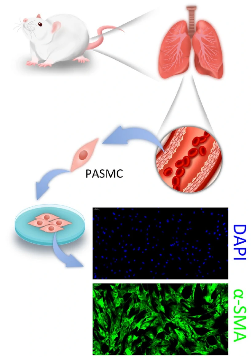
Cells are negative for bacteria, yeast, fungi, and mycoplasma and are characterized by immunofluorescent staining with antibodies to α-smooth muscle actin.
Cells can be expanded on a multiwell culture plate ready for experiments under the cell culture conditions specified by Creative Bioarray.
Repeated freezing and thawing of cells is not recommended.
Standard biochemical procedures performed with smooth muscle cell cultures include the assays of cell to cell interaction, PCR, Western blotting, immunoprecipitation, immunofluorescent staining, immunofluorescent flow cytometry or generating cell derivatives for desired research applications.
Rat Pulmonary Artery Smooth Muscle Cells (RPASMCs) are one of many cell types that comprise the pulmonary arterial system. As cells found in the medial layer of the pulmonary artery, RPASMCs maintain vascular tone and provide structural support. Pulmonary smooth muscle cells are highly plastic and transition between a "contractile phenotype" (regulating blood pressure) and "synthetic phenotype" (repairing/remodeling tissue). When cultured, they display a "hill-and-valley" pattern of proliferation and express cell surface markers such as α-smooth muscle actin (α-SMA) and smooth muscle myosin heavy chain (SM-MHC).
RPASMCs are often used to model Pulmonary Arterial Hypertension (PAH). Pulmonary artery smooth muscle cells are usually cited in papers examining possible molecular causes of PAH including cues that trigger vascular remodeling events such as hyper-proliferation and apoptosis-resistance during hypoxic exposures. Papers targeting HIF-signaling and dysregulation of Kv channels within PASMCs cause pulmonary vasoconstriction.
RPASMCs can also be used to study mechanotransduction from stretching and for high-throughput drug screening. Primary RPASMCs are the preferred in vitro assay to study calcium signaling and vasodilatory drugs because their responses best emulate the response of an intact rat lung before in vivo animal models are utilized.
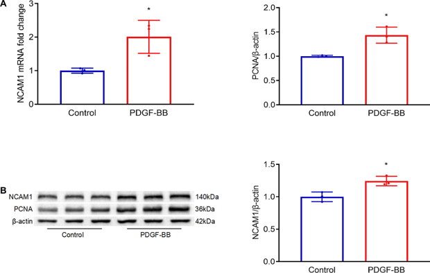
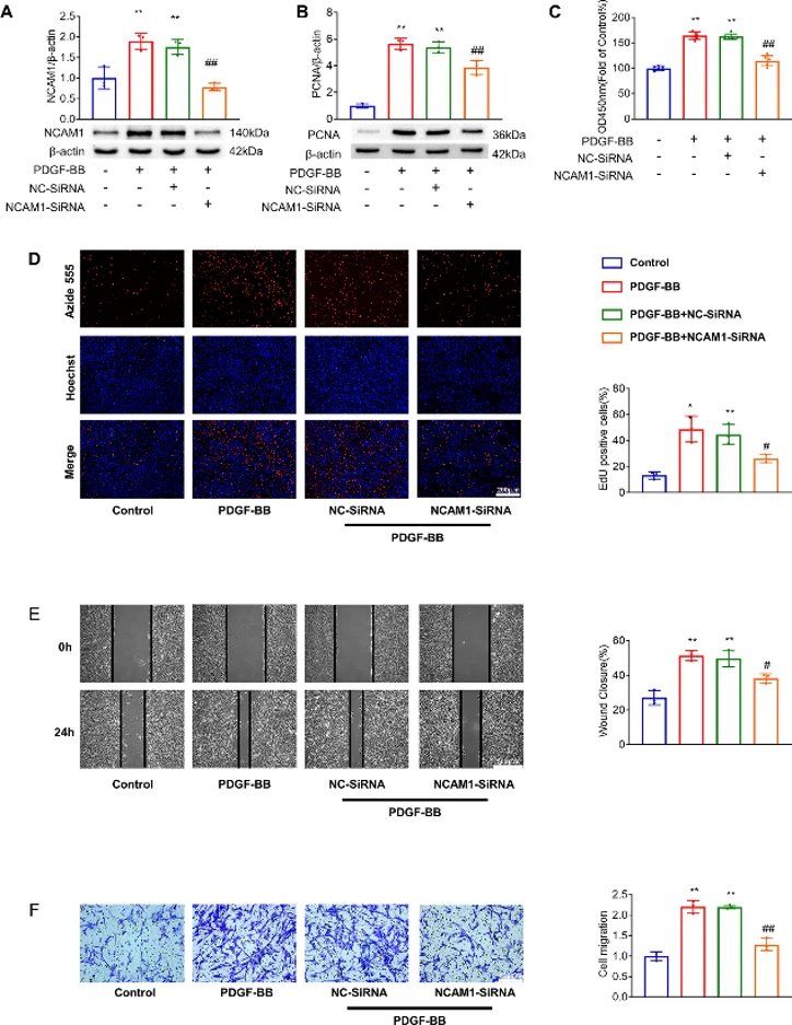
NCAM1 Contributes to the Proliferation and Migration of PDGF-BB-Induced PASMCs
Pulmonary hypertension (PH) involves pulmonary arterial remodeling. Neural cell adhesion molecule 1 (NCAM1) is a cell surface glycoprotein that participates in diverse diseases such as cardiovascular disease, but its role in PH remains unclear. Herein, Chen's team examined the role of NCAM1 in PDGF-BB-stimulated rat pulmonary artery smooth muscle cells (PASMCs). NCAM1 expression was significantly increased in PDGF-BB-stimulated PASMCs at the mRNA level (Fig. 1A) and protein level (Fig. 1B). To determine the roles of NCAM1, they knocked down NCAM1 expression with siRNA (Fig. 2A). NCAM1 knockdown partially inhibited PDGF-BB-induced PCNA increase (Fig. 2B) and suppressed PDGF-BB-induced cell proliferation (Fig. 2C and D) as assessed by CCK-8 assay and EdU incorporation assay. Moreover, NCAM1 knockdown inhibited PDGF-BB-induced PASMC migration (Fig. 2E, F).
Ask a Question
Write your own review

Description: The thoracic aorta is located in the chest cavity and gives off arteries that branch to the esophagus, pericardium, lungs, and trachea. The thoracic aorta can be subdivided into the ascending aorta, ...

Description: Rat Podocytes are isolated from normal rat kidney. The cells are characterized by immunofluorescence with antibodies specific to podocin, Ang1, Nephrin, ACTN4, NPHS2. T25 flasks is required for cell ...

Description: Rat Bronchial Smooth Muscle Cells are isolated from normal rat bronchi tissue. Rat Bronchial Smooth Muscle Cells are characterized by immunofluorescence with antibodies specific to alpha-actin. T25 ...

Description: Guinea Pig Endothelial Cells from Creative Bioarray are isolated from guinea pig tissue. Prior to shipping, cells at passage 2 are detached from flasks and immediately cryopreserved in vials. Each ...

Description: Rat Lung Epithelial Cells are isolated from normal rat lung tissue. The cells are characterized by immunofluorescence with antibodies specific to CK-18, CK-19. T25 flasks is required for cell ...

Description: Rat Vein Endothelial Cells from Creative Bioarray are isolated from inferior vena cava tissue of 6-8 week old laboratory Sprague-Dawley rat. Rat Vein Endothelial Cells are grown in T75 tissue culture ...