
Rat Primary Thyroid Epithelial Cells
Cat.No.: CSC-C4145X
Species: Rat
Source: Thyroid
Cell Type: Epithelial Cell
- Specification
- Background
- Scientific Data
- Q & A
- Customer Review
Rat Thyroid Epithelial Cells are characterized by immunofluorescent staining with antibodies of E-cadherin and ZO-1. Rat Thyroid Epithelial Cells are negative for bacteria, yeast, fungi, and mycoplasma. Cells can be expanded for 3-7 passages at a split ratio of 1:2 or 1:3 under the cell culture conditions specified by Creative Bioarray. Repeated freezing and thawing of cells is not recommended.
Standard biochemical procedures performed with epithelial cell cultures include the assays of cell to cell adhesion and migration, RT-PCR, Western blotting, immunoprecipitation, immunofluorescent staining or immunofluorescent flow cytometry or generating cell derivatives for desired research applications.
Rat Primary Thyroid Epithelial Cells are epithelial cells, one of the most common types of specialized cells that have been isolated from tissues of the thyroid gland of rats. Thyroid epithelial cells are responsible for creating, storing, and secreting hormones produced by the thyroid gland, thyroxine and triiodothyronine. As these are primary thyroid cells rather than immortalized thyroid cell lines, they retain much of the structural and functional differentiation that is found in normal thyroid cells in vivo.
Rat Primary Thyroid Epithelial Cells grow as typical epithelial cells in culture, generally showing a cobblestone shape. When cultured on a suitable extracellular matrix and given Thyroid Stimulating Hormone (TSH), these cells will form spherical follicles. They express several biomarkers that are used to identify thyroid cells including thyroglobulin (Tg), thyroid peroxidase (TPO), and sodium-iodide symporter (NIS). These cells are widely utilized in pharmacological and toxicological studies to evaluate the impact of environmental disruptors on endocrine function.
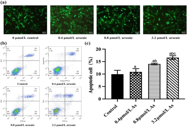
Effects of Arsenic Exposure in Rat Thyroid Cells
Arsenic-induced disease may be related to toxicity to the thyroid gland and endocrine system, however, how this occurs remains unknown. Ma et al. examined whether PI3K and Nrf2 pathways were involved in arsenic-induced toxicity by exposing rats to sodium arsenite (NaAsO2) and rat thyroid epithelial cells. Under light microscopy, thyroid cells treated with vehicle solution in vitro adhered well to the surface of culture wells, were evenly distributed, and showed clear edges (Fig. 1a, upper-left panel). Thyroid cells in the low-exposure group grew densely. In the medium-exposure group, some thyroid cells shrank with unclear edges, and no proliferation was observed. Edges and nuclear staining of cells became blurred in the high-exposure group and gaps between cells were observed (Fig. 1a). Flow cytometry demonstrated significant increases in the apoptotic population of thyroid cells in the medium- and high-exposure groups compared with the low-exposure group (Fig. 1b). The higher the arsenic concentration, the higher the apoptotic rate became, and the high-As group had the highest percentage of apoptotic cells (Fig. 1c).
Ask a Question
Write your own review

Description: The thoracic aorta is located in the chest cavity and gives off arteries that branch to the esophagus, pericardium, lungs, and trachea. The thoracic aorta can be subdivided into the ascending aorta, ...

Description: Rat Podocytes are isolated from normal rat kidney. The cells are characterized by immunofluorescence with antibodies specific to podocin, Ang1, Nephrin, ACTN4, NPHS2. T25 flasks is required for cell ...

Description: Rat Bronchial Smooth Muscle Cells are isolated from normal rat bronchi tissue. Rat Bronchial Smooth Muscle Cells are characterized by immunofluorescence with antibodies specific to alpha-actin. T25 ...

Description: Guinea Pig Endothelial Cells from Creative Bioarray are isolated from guinea pig tissue. Prior to shipping, cells at passage 2 are detached from flasks and immediately cryopreserved in vials. Each ...

Description: Rat Lung Epithelial Cells are isolated from normal rat lung tissue. The cells are characterized by immunofluorescence with antibodies specific to CK-18, CK-19. T25 flasks is required for cell ...

Description: Rat Vein Endothelial Cells from Creative Bioarray are isolated from inferior vena cava tissue of 6-8 week old laboratory Sprague-Dawley rat. Rat Vein Endothelial Cells are grown in T75 tissue culture ...