
Human Pulmonary Fibroblasts
Cat.No.: CSC-C9363W
Species: Human
Source: Lung
Morphology: Multipolar
Cell Type: Fibroblast
- Specification
- Background
- Scientific Data
- Q & A
- Customer Review
Human Pulmonary Fibroblasts (HPFs) are primary fibroblast cells isolated from human lung tissue. Pulmonary fibroblasts are one of the primary structural cell types that maintain the pulmonary interstitium architecture. The cells are characterized by an elongated spindle morphology and robust extracellular matrix production (collagen and fibronectin, amongst others), providing mechanical support to the alveoli and airways. HPFs are specialized in active sensing and responses to stimuli of the lung microenvironment such as mechanical stretch, hypoxia, or inflammatory cues.
HPFs readily undergo activation (phenotypic transition) upon injury related or profibrotic stimuli, leading to enhanced matrix production and contractility. While this serves a physiological role in maintaining normal lung homeostasis and repair after acute lung injury, the mechanisms become dysregulated in disease states, such as pulmonary fibrosis, leading to excessive tissue remodeling and pathology. In this sense, HPFs are frequently used to study lung remodeling, fibrosis, and chronic inflammatory processes. Furthermore, the robust cytokine and growth factor responsiveness in HPFs make them a popular cell line for in vitro studies of fibroblast activation, cell-matrix interactions, and evaluation of antifibrotic or anti-inflammatory compounds in a lung-specific cellular context.
Tracing Micro and Nanoplastics Toxicity in Human Pulmonary Fibroblasts through Integrated Raman and Transcriptomic Analyses
Inhaled micro- and nanoplastics can reach the lungs' distal regions, where their elimination is limited. This study investigated the impact of primary polystyrene micro- and nanoparticles on human pulmonary fibroblasts, focusing on size-dependent internalization and cellular responses to assess potential health risks.
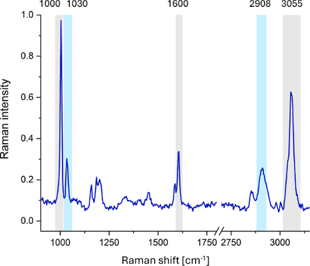
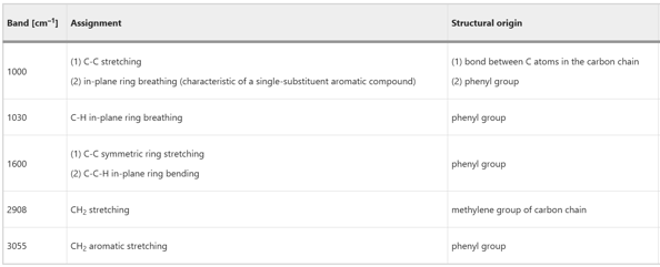
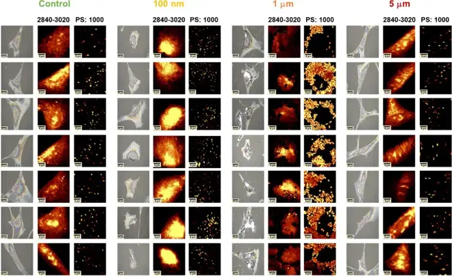
To check if MNPs can get inside HPFs, Raman microscopy was used. Figure 1 shows key Raman bands for PS at around 1000, 1030, 1600, 2908, and 3055 cm⁻¹, with origins detailed in Figure 2. Figure 3 compares topographic analysis of the 1000 cm⁻¹ band (indicating PS) with chemical maps of the 2840-3020 cm⁻¹ region. The 1000 cm⁻¹ band relates to aromatic compounds, while higher wavenumbers show cellular organic matter. For HPFs exposed to 5 μm particles, no significant differences were seen compared to controls. However, HPFs exposed to 1 μm particles clearly showed MNPs in chemical maps, with increased Raman signals at 1000 cm⁻¹ and decreased cellular signals. Despite this, it's unclear if the particles were internalized. Depth scans also couldn't confirm particle penetration due to cell drying, which thinned the cells too much. Future work might involve measuring cells in an aqueous environment to address this.
When thawing frozen tubes out of the liquid nitrogen tank, safety must be observed and protective glasses and gloves can be worn to prevent bursting of the tubes. Immediately after removing the tubes, put them into a 37℃ water bath for rapid thawing. Shake the tubes gently so that they melt completely within 1 minute to avoid damage to the cells due to intracellular recrystallization caused by the penetration of water into the cells as a result of slow thawing. Be careful that the water surface does not exceed the edge of the freezing tube lid, otherwise contamination may occur.
Ask a Question
Average Rating: 5.0 | 1 Scientist has reviewed this product
High-qulity products
The high quality of the products with favourable prices has enabled us to establish a long-term relationship.
25 Jan 2022
Ease of use
After sales services
Value for money
Write your own review
Description: Human Pulmonary Artery Fibroblasts are isolated from normal human pulmonary artery.
Description: Primary Human Fetal Lung Fibroblast Cells were initiated from human lung tissue (normal 25 week gestation).These cells were originated using Complete Serum-Free Medium Kit With SuperFuel™, are ...

Description: GFP-Human Lung Airway Smooth Muscle Cells (GFP-HLAWSMCs) provided by Creative Bioarray are puromycin-selected from primary human lung airway smooth muscle cells infected with lentivirus expressing ...

Description: ACE2-GFP Expressing Human Pulmonary Artery Endothelial Cells (ACE2-GFP-HPAECs) provided by Creative Bioarray are puromycin-selected from Human Pulmonary Artery Endothelial Cells infected with ...

Description: HTSMC from Creative Bioarray Research are isolated from human trachea. HTSMC are cryopreserved at secondary culture and delivered frozen. Each vial contains >5 x 10^5 cells in 1 ml volume. HTSMC are ...
Description: The human type 1 alveolar epithelial cells from Creative Bioarray are isolated from human alveolar tissue. These cells are shipped at passage 2 and maintained in SuperCult® human type 1 alveolar ...