DX3
Cat.No.: CSC-C9373L
Species: Homo sapiens (Human)
Source: Hypodermis Metastasis
Culture Properties: monolayer
- Specification
- Background
- Scientific Data
- Q & A
- Customer Review
Shipping Condition: Room Temperature
The DX3 cell line is a highly characterized human melanoma model derived from a metastatic site, offering researchers a robust platform for investigating the molecular mechanisms of tumor progression, epithelial-mesenchymal transition (EMT), and pharmacological resistance. As melanoma remains one of the most aggressive malignancies, the DX3 line serves as a critical tool for bridging the gap between basic oncogenic signaling research and preclinical drug development.
- Stable Metastatic Phenotype: Unlike early-stage primary melanoma lines, DX3 cells exhibit a stabilized metastatic profile. This makes them exceptionally suited for migration and invasion assays, as well as in vivo xenograft models to study organ-specific colonization.
- Well-Defined Genetic Background: Our DX3 cells are rigorously authenticated via STR profiling and characterized for key melanoma drivers. Their distinct mutational status provides a reliable baseline for studying MAPK pathway signaling (BRAF/MEK) and evaluating the efficacy of targeted kinase inhibitors.
- High Transfection Efficiency & Plasticity: DX3 cells demonstrate superior amenability to genetic manipulation, including CRISPR/Cas9 gene editing and lentiviral transduction. This plasticity allows for the rapid generation of reporter lines or knockout models to identify novel therapeutic targets.
- Optimized for High-Throughput Screening (HTS): With a consistent doubling time and predictable growth kinetics in standard DMEM/RPMI media, DX3 is an ideal candidate for large-scale small molecule libraries and toxicity profiling, ensuring high reproducibility across multi-well formats.
By incorporating the DX3 cell line into your oncology pipeline, you leverage a clinically relevant human model that provides the physiological depth necessary for high-impact cancer research and drug validation.
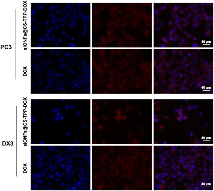
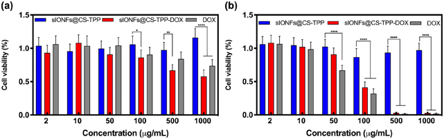
In Vitro Cellular Uptake and Cytotoxicity Study of pH-Responsive Magnetic Iron Oxide Nanoflower-Chitosan Nanogels
There has been a recent surge in the development of drug delivery systems that are specifically targeted towards tumors. The primary objective of these systems is to enhance the efficacy of anti-tumor drugs while simultaneously reducing any potential harmful side effects. This study focuses on designing a surface coating that is pH-responsive for iron oxide nanoflower (IONF) cores, allowing the controlled release of the drug doxorubicin (DOX). To achieve this, IONFs were coated with chitosan (CS) using NaOH, tripolyphosphate (TPP), and glutaraldehyde (GLU) as crosslinking agents. The biocompatibility of the developed formulation was investigated through the MTT assay, which revealed the safety of the formulation in biological systems. Finally, flow cytometry analysis was performed to evaluate the efficiency of the drug delivery system.
Ask a Question
Write your own review
- You May Also Need

Description: Established from the primary tumor (right cervical) of a 64-year-old woman with cutaneous melanoma

Description: Established from the lymph node metastasis of a malignant melanoma from a 42-year-old Caucasian woman

Description: Species: human - male, 31 years old, CaucasianIsoenzyme: G6PD, BProduction: melaninHistopathology: melanoma
Description: Epstein-Barr virus-positive cell line established from peripheral blood in 1977 from male patient with melanoma

Description: Established from the primary tumor of a 58-year-old woman with melanoma in 1977

Description: Established from the primary (achromic) cutaneous tumor (left thigh) of a 26-year-old man with malignant melanoma (primary tumor histology: SSM level IV); same patient as cell line IGR-37; described ...
- Adipose Tissue-Derived Stem Cells
- Human Neurons
- Mouse Probe
- Whole Chromosome Painting Probes
- Hepatic Cells
- Renal Cells
- In Vitro ADME Kits
- Tissue Microarray
- Tissue Blocks
- Tissue Sections
- FFPE Cell Pellet
- Probe
- Centromere Probes
- Telomere Probes
- Satellite Enumeration Probes
- Subtelomere Specific Probes
- Bacterial Probes
- ISH/FISH Probes
- Exosome Isolation Kit
- Human Adult Stem Cells
- Mouse Stem Cells
- iPSCs
- Mouse Embryonic Stem Cells
- iPSC Differentiation Kits
- Mesenchymal Stem Cells
- Immortalized Human Cells
- Immortalized Murine Cells
- Cell Immortalization Kit
- Adipose Cells
- Cardiac Cells
- Dermal Cells
- Epidermal Cells
- Peripheral Blood Mononuclear Cells
- Umbilical Cord Cells
- Monkey Primary Cells
- Mouse Primary Cells
- Breast Tumor Cells
- Colorectal Tumor Cells
- Esophageal Tumor Cells
- Lung Tumor Cells
- Leukemia/Lymphoma/Myeloma Cells
- Ovarian Tumor Cells
- Pancreatic Tumor Cells
- Mouse Tumor Cells