
- Specification
- Background
- Scientific Data
- Q & A
- Customer Review
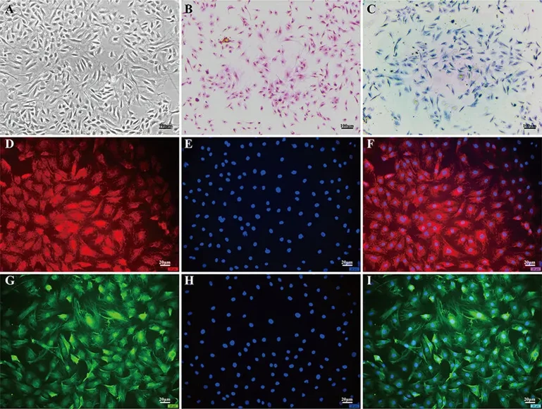
Mouse nucleus pulposus cells from Creative Bioarray are isolated from the mouse intervertebral disc tissue. The method we use to isolate mouse nucleus pulposus cells was developed based on a combination of established and our proprietary methods. The mouse nucleus pulposus cells are characterized by immunofluorescence with antibodies specific to collagen II.Each vial contains 0.5x10^6 cells per ml and is delivered frozen.
Mouse nucleus pulposus (NP) cells are intervertebral disc cells located in the center of the disc. NP cells are mesenchymal in origin and descend from notochordal precursors during development, though mouse NP cells are known to undergo changes to a more chondrocyte-like phenotype in adult tissues. Mouse NP cells have been used as a model system for human disc degeneration. These cells are rounded or oval in shape and have an extracellular matrix with high concentrations of proteoglycans and type II collagen.
In vitro, mouse NP cells display limited proliferative capacity and are sensitive to environmental cues, requiring hypoxic culture conditions and supplementation with growth factors such as TGF-β to preserve their native phenotype. Functionally, these cells regulate extracellular matrix synthesis, sense mechanical loading, and modulate inflammatory responses. Under degenerative or injury conditions, NP cells upregulate pro-inflammatory cytokines and matrix-degrading enzymes, contributing to tissue breakdown and pain-associated pathways.
Mouse NP cells have been used in various areas of intervertebral disc research including the study of degenerative mechanisms, disc mechanobiology, regenerative therapies, and biomaterial testing. These cells can also be integrated into transgenic or knockout mouse lines to study molecular signaling pathways in intervertebral disc homeostasis and degeneration.
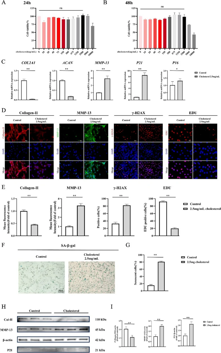
High Cholesterol can Directly Promote Metabolic Disorders in NPCs
Intervertebral disc degeneration (IVDD) is a major cause of low back pain (LBP). Dyslipidaemia can induce chronic inflammation, promote macrophage polarization, and may affect intervertebral disc (IVD) homeostasis. However, the relationship between dyslipidaemia and IVDD is unclear.
In this retrospective study, the baseline characteristics of 196 subjects were included and high TC, TG and LDL-C were identified as independent risk factors for IVDD. TC was selected as the indicator for further experiments. Mouse nucleus pulposus cells (NPCs) were cultured with water-soluble cholesterol. CCK-8 assays were used to evaluate the toxicity to NPCs and there was no significant difference under the concentration of cholesterol 5000 µg/mL at 24 h (Fig. 1A) and 2500 µg/mL at 48 h (Fig. 1B). The Chinese guidelines for lipid management (2023) was searched and hypercholesterolemia was diagnosed with TC > 6.2 mmol/L (~2400 µg/mL). Therefore, 2500 µg/mL of cholesterol was used for further experiments. Compared mRNA levels showed that cholesterol-treated NPCs had lower levels of benign matrix markers (COL2A1 and ACAN) and higher levels of MMP13 (~3 folds) and senescence markers (P21 and P16) (Fig. 1C). Immunofluorescence staining confirmed these results (Fig. 1D, E). γ-H2AX staining, a marker of cellular senescence, demonstrated significant senescence of NPCs under high-cholesterol condition, which is consistent with qPCR and Western blot analysis (Fig. 1D, E). EdU assays showed that NPCs had an inhibited proliferation ability in high-cholesterol condition (Fig. 1D, E). β-Galactosidase staining of NPCs showed a ~4-fold increase of senescent cells under cholesterol treatment (Fig. 1F). Western blotting also confirmed the above results (Fig. 1G). The results indicate that high cholesterol can directly accelerate metabolic disorders in NPCs, promote NPC aging and inhibit NPC proliferation, and finally accelerate IVDD.
Ask a Question
Write your own review

Description: C57BL/6-GFP Mouse Skeletal Muscle Microvascular Endothelial Cells from Creative Bioarray are isolated from C57BL/6-Tg (CAG-EGFP) 1Osb/J mouse skeletal muscle tissue of pathogen-free laboratory mice. ...

Description: eNOS KO Mouse Stomach Epithelial Cells from Creative Bioarray are isolated from stomach tissue of pathogen-free laboratory mice. eNOS KO Mouse Stomach Epithelial Cells are grown in a T25 tissue ...

Description: eNOS KO Mouse Liver Fibroblasts from Creative Bioarray are isolated from liver tissue of pathogen-free laboratory mice. eNOS KO Mouse Liver Fibroblasts are grown in T75 tissue culture flasks ...

Description: C57BL/6-GFP Mouse Corneal Epithelial Cells from Creative Bioarray are isolated from C57BL/6-GFP-Tg(CAG-EGFP)1Osb/J mouse corneal tissue of pathogen-free laboratory mice. C57BL/6-GFP Mouse Corneal ...

Description: BALB/c Mouse Retinal Microvascular Endothelial Cells from Creative Bioarray are isolated from retinal tissue of pathogen-free laboratory mice. BALB/c Mouse Retinal Microvascular Endothelial Cells are ...