mouse lung fibroblasts
Cat.No.: CSC-C1937
Species: Mouse
Source: Lung
Cell Type: Fibroblast
- Specification
- Background
- Scientific Data
- Q & A
- Customer Review
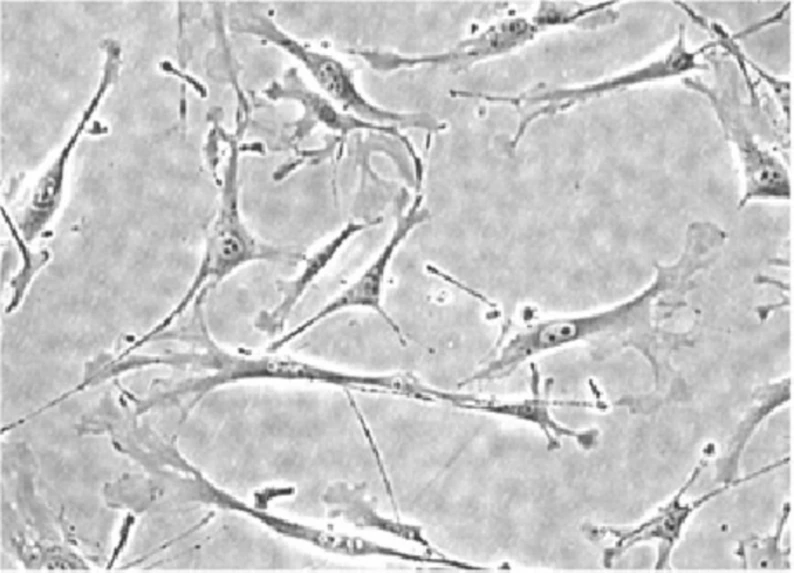
Mouse lung fibroblasts are lung stromal cells of the interstitial and alveolar compartments in the mouse. These adherent spindle-shaped cells continuously synthesize and remodel the extracellular matrix to maintain the structural integrity of the lung. In the healthy lung, they are important in maintaining alveolar stability and contribute to wound repair, as well as regulating tissue elasticity. Upon activation through cytokines such as TGF-β, lung fibroblasts can undergo FMT, an α-SMA+ phenotype, and produce excessive ECM, which is the basis for lung fibrosis.
Primary mouse lung fibroblasts, as well as immortalized cell lines, are used extensively in studies of respiratory diseases. These cells are used to understand the molecular underpinnings of idiopathic pulmonary fibrosis, acute lung injury, and virus-induced inflammation. They can also be used to study fibroblast-immune cell communication by secreting cytokines like IL-6, TNF-α, and CXCL1. Mouse lung fibroblasts can also be used to model tumor-stroma interactions in the lung and cancer-associated fibroblast activation. Mouse lung fibroblasts are also used in drug screens for antifibrotic and anti-TGF-β agents, ECM remodeling modulators, and many other lung-relevant drugs. Lung fibroblasts have also been used in the modeling of ventilation-induced injury due to their response to mechanical stress.
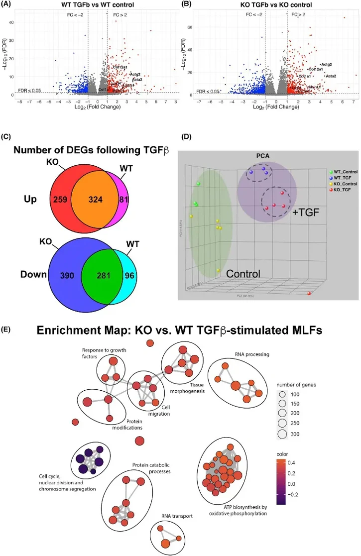
Tollip-/- Mouse Lung Fibroblasts Exhibit Enhanced Response to TGFβ
Variations in the TOLLIP gene correlate with disease risk and therapy response in idiopathic pulmonary fibrosis. Here, Chow et al. stimulated primary mouse lung fibroblasts (MLFs) with TGFβ and assessed transcriptional and functional responses.
TOLLIP overexpression in HEK293T and HepG2 cells reduces TGFβ responsiveness, suggesting TOLLIP may modulate TGFβ signaling. RNA-seq analysis of WT and Tollip-/- mouse lung fibroblasts (MLFs) treated with TGFβ showed that both upregulated myofibroblast differentiation genes (Fig. 1A, B), but KO MLFs had significantly more differentially expressed genes (Fig. 1A, B). WT MLFs had 405 upregulated and 377 downregulated genes, while Tollip-/- MLFs had 583 upregulated and 671 downregulated genes (Fig. 1C). Principal component analysis revealed a strong effect of TGFβ on MLF transcriptional signatures (Fig. 1D), with Tollip-/- MLFs showing greater responsiveness to TGFβ stimulation.
Ask a Question
Write your own review

Description: C57BL/6-GFP Mouse Skeletal Muscle Microvascular Endothelial Cells from Creative Bioarray are isolated from C57BL/6-Tg (CAG-EGFP) 1Osb/J mouse skeletal muscle tissue of pathogen-free laboratory mice. ...

Description: eNOS KO Mouse Stomach Epithelial Cells from Creative Bioarray are isolated from stomach tissue of pathogen-free laboratory mice. eNOS KO Mouse Stomach Epithelial Cells are grown in a T25 tissue ...
Description: eNOS KO Mouse Liver Fibroblasts from Creative Bioarray are isolated from liver tissue of pathogen-free laboratory mice. eNOS KO Mouse Liver Fibroblasts are grown in T75 tissue culture flasks ...
Description: C57BL/6-GFP Mouse Corneal Epithelial Cells from Creative Bioarray are isolated from C57BL/6-GFP-Tg(CAG-EGFP)1Osb/J mouse corneal tissue of pathogen-free laboratory mice. C57BL/6-GFP Mouse Corneal ...

Description: BALB/c Mouse Retinal Microvascular Endothelial Cells from Creative Bioarray are isolated from retinal tissue of pathogen-free laboratory mice. BALB/c Mouse Retinal Microvascular Endothelial Cells are ...