
Mouse Lens Epithelial Cells
Cat.No.: CSC-C5407S
Species: Mouse
Source: Lens; Eye
Cell Type: Epithelial Cell
- Specification
- Background
- Scientific Data
- Q & A
- Customer Review
Mouse lens epithelial cells from Creative Bioarray are isolated from the mouse eye tissue. The method we use to isolate mouse lens epithelial cells was developed based on a combination of established and our proprietary methods. The mouse lens epithelial cells are characterized by immunofluorescence with antibodies specific to cytokeratin-18 (CK-18). Each vial contains 0.5x10^6 cells per ml and is delivered frozen.
Mouse Lens Epithelial Cells (mLECs) are a single layer of cuboidal cells, positioned immediately underneath the anterior capsule of the lens. They form the major source of progenitors for the entire avascular and transparent lens organ. These cells are characterised by a spatially-regulated differentiation program in which while the central cells are quiescent, cells at the equatorial region of the lens undergo proliferation, and also activate an impressive differentiation trajectory: mLECs undergo dramatic elongation, followed by organelle degradation and synthesis of enormous amounts of crystallins, leading to their differentiation into anucleate lens fibres.
mLECs play a key role in supporting the growth and homeostasis of the lens throughout life. These cells form the main site for the antioxidant response and metabolic activity of the lens, playing an important role in protection against oxidative damage, which is a major factor in cataract development. These cells also precisely regulate ion and osmotic homeostasis to prevent light-scattering and opacification of the lens. Primary and immortalized lines of mLECs (αTN4-1) are frequently used to study the mechanisms of cataract formation, lens development/regeneration and to screen potential therapeutics/toxins on ocular tissues in vitro.
CREB Regulates Expression of Mesenchymal Genes in Lens Epithelial Cells
Epithelial mesenchymal transition (EMT) of lens epithelial cells (LECs) is crucial in lens fibrotic disorders, but its regulation is not fully understood. Here, Zhang's team explored CREB's role in lens EMT using mouse LECs and an anterior subcapsular cataract model.
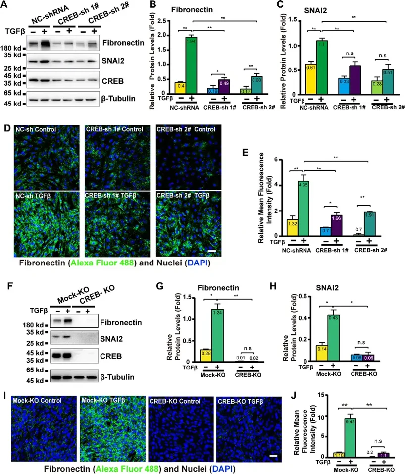
To understand CREB's role in the ocular lens, they examined CREB protein expression in 2-month-old mouse ocular tissues (retina, cornea, lens epithelium) and major organs (heart, liver, lung, brain, muscle). CREB was moderately expressed in lens epithelium, lower than in cornea and lung, and remained stable at postnatal 1, 2, and 6 months. Previous studies identified CREB-binding sites in the core promoters of mesenchymal marker genes Fn1 and Snai2. EMSA showed that nuclear extracts from mouse lens epithelial cells specifically bound to Fn1 and Snai2 probes, confirming CREB binding to these promoters. This suggests CREB regulates mesenchymal genes. Given TGFβ-induced EMT's link to posterior capsular opacification, they knocked down CREB in mouse lens epithelial cells and found that TGFβ-induced Fibronectin and SNAI2 expression was significantly reduced (Fig. 1A-C). In CREB knockout (CREB-KO) cells, TGFβ treatment failed to induce Fibronectin and SNAI2 expression (Fig. 1F-H). Transfecting CREB-KO cells with WT-CREB partially restored these proteins. These results show CREB is a key transcription factor controlling lens epithelial cell EMT.
Ask a Question
Write your own review
- You May Also Need


Description: C57BL/6-GFP Mouse Skeletal Muscle Microvascular Endothelial Cells from Creative Bioarray are isolated from C57BL/6-Tg (CAG-EGFP) 1Osb/J mouse skeletal muscle tissue of pathogen-free laboratory mice. ...

Description: eNOS KO Mouse Stomach Epithelial Cells from Creative Bioarray are isolated from stomach tissue of pathogen-free laboratory mice. eNOS KO Mouse Stomach Epithelial Cells are grown in a T25 tissue ...

Description: eNOS KO Mouse Liver Fibroblasts from Creative Bioarray are isolated from liver tissue of pathogen-free laboratory mice. eNOS KO Mouse Liver Fibroblasts are grown in T75 tissue culture flasks ...
Description: C57BL/6-GFP Mouse Corneal Epithelial Cells from Creative Bioarray are isolated from C57BL/6-GFP-Tg(CAG-EGFP)1Osb/J mouse corneal tissue of pathogen-free laboratory mice. C57BL/6-GFP Mouse Corneal ...

Description: BALB/c Mouse Retinal Microvascular Endothelial Cells from Creative Bioarray are isolated from retinal tissue of pathogen-free laboratory mice. BALB/c Mouse Retinal Microvascular Endothelial Cells are ...