
Mouse Extrahepatic Bile Duct Epithelial Cells
Cat.No.: CSC-C5309S
Species: Mouse
Source: Bile Duct
Cell Type: Epithelial Cell; Cholangiocyte
- Specification
- Background
- Scientific Data
- Q & A
- Customer Review
Mouse extrahepatic bile duct epithelial cells from Creative Bioarray are isolated from the mouse bile duct tissue. The method we use to isolate mouse extrahepatic bile duct epithelial cells was developed based on a combination of established and our proprietary methods. The mouse extrahepatic bile duct epithelial cells are characterized by immunofluorescence with antibodies specific to pan-cytokeratin (PCK). Each vial contains 0.5x10^6 cells per ml and is delivered frozen.
Mouse Extrahepatic Bile Duct Epithelial Cells (EHBDECs) are primary cholangiocytes isolated from the extrahepatic bile ducts of mice. These epithelial cells line the extrahepatic bile ducts and contribute to the structural and functional integrity of the bile ducts. They serve as a cellular model in vitro for the study of biliary epithelial biology in a controlled experimental environment.
Mouse EHBDECs are commonly utilized in investigations of bile duct development, epithelial homeostasis, inflammatory responses and tissue repair. They can be used to study cholangiocyte proliferation, epithelial barrier function, cell-cell communication, and signaling pathways in biliary physiology. In recent years, extrahepatic bile duct epithelial cells have been incorporated into bile duct organoid systems and experimental models of biliary injury to investigate cellular responses during regeneration and disease progression.
These cells serve as an in vitro model to investigate cholangiopathies such as biliary atresia, bile duct obstruction and biliary fibrosis. They can also be used to assess molecular mechanisms associated with pathways such as Wnt, Notch, TGF-β and inflammatory signaling. Mouse Extrahepatic Bile Duct Epithelial Cells are a primary cell model isolated from native tissue and can be used to support studies of biliary epithelial function and provide experimental data for hepatobiliary research.
Canonical WNT Signaling Drives Cholangiocyte Hyperproliferation in Extrahepatic Bile Duct Obstruction
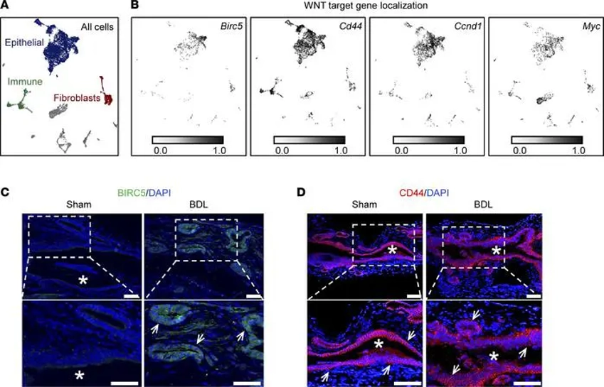
Extrahepatic bile duct (EHBD) obstruction leads to cholangiocyte hyperproliferation, a key feature of severe cholangiopathies. To investigate the role of WNT signaling, Calder et al. analyzed WNT target gene expression in sham and bile duct ligation (BDL) models. Birc5, Cd44, Ccnd1, and Myc were localized primarily to epithelial cell clusters in the EHBD of both conditions (Fig. 1A-B). Immunofluorescence confirmed BIRC5 and CD44 protein expression specifically within epithelial cells (Fig. 1C-D), indicating that cholangiocytes both produce WNT ligands and act as WNT-responsive cells.
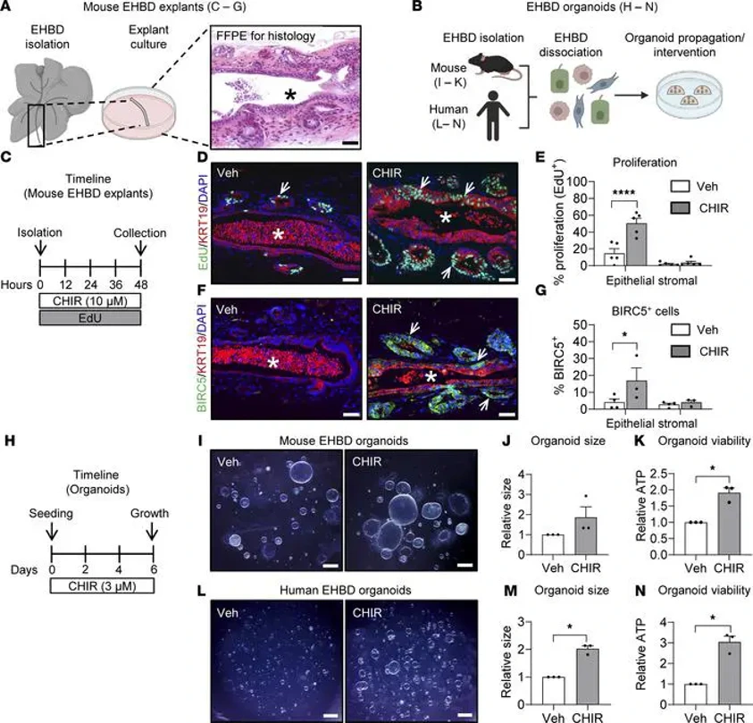
Functional validation was performed using EHBD explant (Fig. 2A) and organoid (Fig. 2B) models. Under homeostatic conditions, explants exhibited low basal proliferation (Fig. 2D). Stimulation with CHIR-99021 (CHIR), a GSK3β inhibitor that activates canonical WNT signaling (Fig. 2C), significantly increased proliferation of KRT19-positive cholangiocytes, as shown by EdU incorporation (Fig. 2D-E). CHIR treatment also upregulated the WNT target protein BIRC5 (Fig. 2F-G), mirroring the in vivo expression pattern observed after BDL. These findings demonstrate that autocrine canonical WNT signaling directly drives obstruction-induced cholangiocyte hyperproliferation.
Ask a Question
Write your own review
- You May Also Need
Description: C57BL/6-GFP Mouse Skeletal Muscle Microvascular Endothelial Cells from Creative Bioarray are isolated from C57BL/6-Tg (CAG-EGFP) 1Osb/J mouse skeletal muscle tissue of pathogen-free laboratory mice. ...
Description: eNOS KO Mouse Stomach Epithelial Cells from Creative Bioarray are isolated from stomach tissue of pathogen-free laboratory mice. eNOS KO Mouse Stomach Epithelial Cells are grown in a T25 tissue ...
Description: eNOS KO Mouse Liver Fibroblasts from Creative Bioarray are isolated from liver tissue of pathogen-free laboratory mice. eNOS KO Mouse Liver Fibroblasts are grown in T75 tissue culture flasks ...
Description: C57BL/6-GFP Mouse Corneal Epithelial Cells from Creative Bioarray are isolated from C57BL/6-GFP-Tg(CAG-EGFP)1Osb/J mouse corneal tissue of pathogen-free laboratory mice. C57BL/6-GFP Mouse Corneal ...
Description: BALB/c Mouse Retinal Microvascular Endothelial Cells from Creative Bioarray are isolated from retinal tissue of pathogen-free laboratory mice. BALB/c Mouse Retinal Microvascular Endothelial Cells are ...