
Immortalized Rat Hepatocytes-SV40
Cat.No.: CSC-I9161L
Species: Rattus norvegicus
Source: Liver
Morphology: Polyhedral
Culture Properties: Adherent
- Specification
- Background
- Scientific Data
- Q & A
- Customer Review
Note: Never can cells be kept at -20 °C.
CIK-HT003 HT® Lenti-SV40T Immortalization Kit
Immortalized rat hepatocyte cell lines generated via SV40 large T antigen are well-characterized in vitro models for studies in liver biology, drug metabolism, and toxicology.
The primary advantage of Immortalized Rat Hepatocytes-SV40 lies in their unlimited proliferative capacity combined with maintained hepatic differentiation functions. Unlike primary rat hepatocytes which rapidly lose liver‑specific characteristics upon culture, these cells retain measurable activities of phase I (cytochrome P450) and phase II drug‑metabolizing enzymes, with demonstrated inducibility by classic agents including phenobarbital (170% increase for CYPIIB), methylcholanthrene (500% increase for CYPIA), and dexamethasone.
Additional advantages include experimental reproducibility, high scalability, and absence of donor‑to‑donor variability that plagues primary hepatocyte preparations. The expression of SV40 large T antigen in these cells can be confirmed by qPCR, and rigorous quality control ensures they are free from mycoplasma, bacteria, and viral contaminants. Taken together, Immortalized Rat Hepatocytes-SV40 provides a reproducible, ethically uncomplicated, and functionally relevant platform for liver disease modeling, toxicogenomics, high‑throughput drug screening, and bioartificial liver support system development.
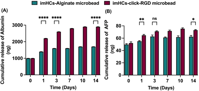
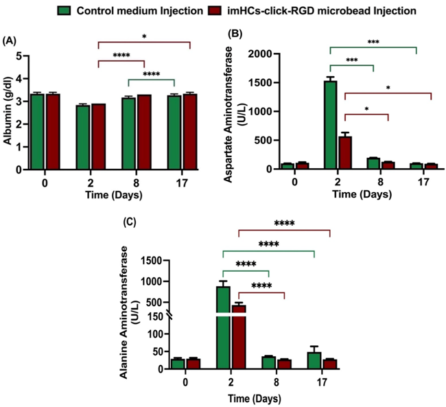
Evaluation of Immortalized Hepatocyte Encapsulated Click-Microbeads with RGD Peptide for Treatment of Liver Failure in Male Rats
Cell encapsulation in biocompatible microbeads offers a promising strategy for cell-based therapy in acute liver failure (ALF). This study evaluates the use of immortalized hepatocyte cells (imHCs) encapsulated in click-arginyl glycyl aspartic acid (click-RGD)-modified alginate microbeads, focusing on their biocompatibility and therapeutic potential. In vitro assessments showed that click-RGD microbeads significantly enhanced cell viability on day 4, spatial distribution, and hepatocyte function, evidenced by increased albumin on day 14 and alpha-fetoprotein (AFP) secretion compared to unmodified alginate microbeads. For in vivo testing, ALF was induced in Sprague-Dawley male rats using D-galactosamine (D-gal), followed by intraperitoneal administration of imHCs-loaded click-RGD microbeads in the treated group and CMRL medium injection in the control group. Treated rats exhibited faster reductions in aspartate aminotransferase (AST) and alanine aminotransferase (ALT) levels, higher albumin production, and improved liver histology, characterized by reduced necrosis and the absence of inflammation, on day 14 after treatment. No adverse host responses were observed, confirming the biocompatibility of the microbeads.
These findings support the potential of click-RGD microbeads as a therapeutic platform for ALF, warranting further studies on long-term implantation, immune response, and co-encapsulation strategies.
Ask a Question
Write your own review

Description: The Immortalized Mouse Prostate Epithelial Cells (iMPEC, clone7) is derived by co-expression of adenovirus E1A and dominant-negative p53 (p53DD). In addition to its non-tumorigenicity, this cell line ...
Description: Immortalized Mouse Primary Artery Fibroblasts-GFP provided by Creative Bioarray have been developed by immortalizing mouse artery fibroblasts with SV40 Large T antigen and transfecting with tGFP. The ...

Description: Immortalized MHC II -/- Mouse Macrophage Cells (C2D) is derived from the knockout mice negative for MHCII molecule. They are stable in culture and survived through crisis in which they spontaneously ...
Description: The bone morphogenetic proteins (BMPs) produced by calvarial cells are crucial in osteoblastic differentiation and bone regeneration, however the limited lifespan of primary cells diminishes their ...
Description: The Immortalized Mouse Atrioventricular Cushion Mesenchymal Cells (tsA58-AVM) were derived from atrioventricular (AV) cushions of H-2Kb -tsA58 embryos at E9.5 and were conditionally immortalized in ...
Description: IDG-SW3 represent a non-homogenous population progressing from early osteoblasts to late osteocytic. These cells express functional SV40 large T antigen that is induced in the presence of IFN γ under ...
- Adipose Tissue-Derived Stem Cells
- Human Neurons
- Mouse Probe
- Whole Chromosome Painting Probes
- Hepatic Cells
- Renal Cells
- In Vitro ADME Kits
- Tissue Microarray
- Tissue Blocks
- Tissue Sections
- FFPE Cell Pellet
- Probe
- Centromere Probes
- Telomere Probes
- Satellite Enumeration Probes
- Subtelomere Specific Probes
- Bacterial Probes
- ISH/FISH Probes
- Exosome Isolation Kit
- Human Adult Stem Cells
- Mouse Stem Cells
- iPSCs
- Mouse Embryonic Stem Cells
- iPSC Differentiation Kits
- Mesenchymal Stem Cells
- Immortalized Human Cells
- Immortalized Murine Cells
- Cell Immortalization Kit
- Adipose Cells
- Cardiac Cells
- Dermal Cells
- Epidermal Cells
- Peripheral Blood Mononuclear Cells
- Umbilical Cord Cells
- Monkey Primary Cells
- Mouse Primary Cells
- Breast Tumor Cells
- Colorectal Tumor Cells
- Esophageal Tumor Cells
- Lung Tumor Cells
- Leukemia/Lymphoma/Myeloma Cells
- Ovarian Tumor Cells
- Pancreatic Tumor Cells
- Mouse Tumor Cells