
Immortalized Rat Hepatic Stellate Cells (HSC-T6)
Cat.No.: CSC-I2673Z
Culture Properties: Adherent
- Specification
- Background
- Scientific Data
- Q & A
- Customer Review
The HSC-T6 cell line is an immortalized rat hepatic stellate cell (HSC) line established by SV40 large T‑antigen transfection of primary stellate cells isolated from an adult male Sprague‑Dawley rat. Hepatic stellate cells are perisinusoidal liver cells specialized in vitamin A (retinoid) storage; upon liver injury they transdifferentiate into activated myofibroblasts, the principal source of extracellular matrix (ECM) in hepatic fibrosis.
The primary advantage of HSC‑T6 is its constitutively activated, myofibroblast‑like phenotype that faithfully recapitulates fibrogenic HSCs without the need for exogenous activation. Unlike quiescent primary HSCs, which spontaneously activate on plastic and exhibit substantial donor‑to‑donor variability, HSC‑T6 provides a homogeneous, reproducible, and unlimited cell supply for mechanistic studies. The cells robustly express fibrotic markers including α‑smooth muscle actin (α‑SMA), glial fibrillary acidic protein (GFAP), and desmin, and secrete TGF‑β, type I collagen, and other ECM components, closely mimicking the activated HSC phenotype in fibrotic liver.
Additional advantages include rapid growth (doubling time ~10 hours), high homogeneity, experimental reproducibility, and the ability to overcome ethical and technical challenges associated with primary HSC isolation-which is time‑consuming, yields low cell numbers, and is subject to significant lot‑to‑lot variation. HSC‑T6 is also amenable to high‑throughput drug screening and has extensive molecular characterization, including a defined short‑tandem‑repeat (STR) profile and transcriptome analysis, ensuring reliable cell authentication. Owing to these features, HSC‑T6 has become a gold‑standard in vitro model for studying hepatic fibrosis, retinoid metabolism, and the pathophysiology of chronic liver diseases such as cirrhosis, as well as for evaluating novel anti‑fibrotic therapies.
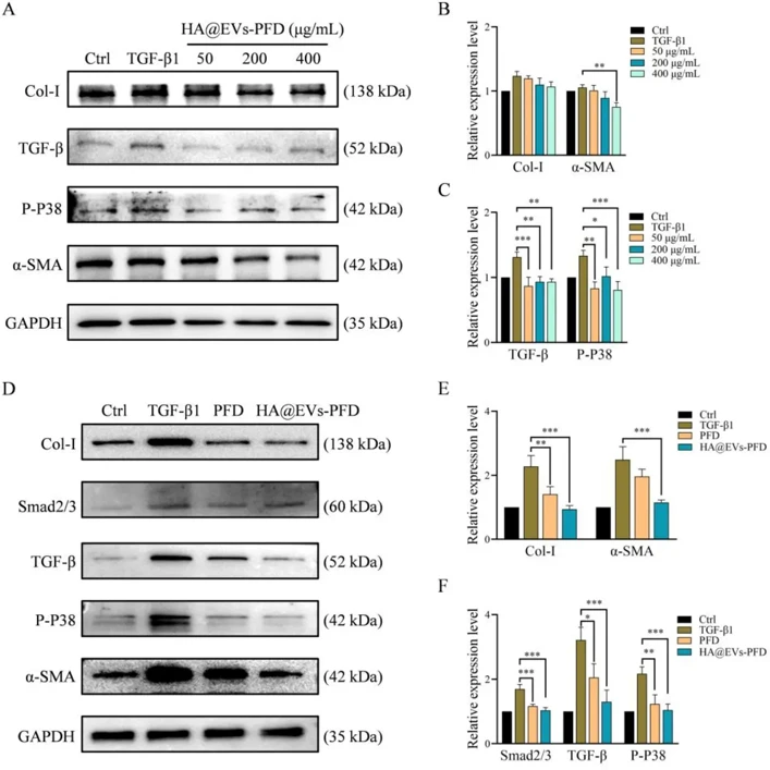
Hyaluronic Acid Modified Extracellular Vesicles Targeting Hepatic Stellate Cells to Attenuate Hepatic Fibrosis
Transforming growth factor-beta1 (TGF-β1) plays a pivotal role in promoting hepatic fibrosis, pirfenidone (PFD) could inhibit TGF-β1 signaling pathway to alleviate hepatic stellate cells (HSC) activation mediated hepatic fibrosis. The targeting delivery strategy of PFD to hepatic stellate cells is a challenge. Extracellular vesicles (EVs), cell-derived membranous particles are intraluminal nano-vesicles that play a vital role in intercellular communication, they also be considered as an ideal nano-carrier.
In this study, we developed a target strategy to deliver PFD to HSC with CD44 over-expression by EVs, hyaluronic acid (HA) modified DSPE-PEG2000 endows the active targeting ability of activated HSCs to PFD-loaded EVs.
In both rat hepatic stellate cell line HSC-T6 and rat hepatocyte cell line BRL, HA@EVs-PFD demonstrated the capacity to down-regulate the expression of collagen-synthesis-related proteins and showed superior inhibition efficacy of HSC-T6 activation compared to free PFD. In hepatic fibrosis model, 4 weeks of HA@EVs-PFD treatment resulted in a reduction in liver collagen fibers, significant improvement in hepatic cell morphology, and amelioration of hepatic fibrosis.
Ask a Question
Write your own review

Description: The Immortalized Mouse Prostate Epithelial Cells (iMPEC, clone7) is derived by co-expression of adenovirus E1A and dominant-negative p53 (p53DD). In addition to its non-tumorigenicity, this cell line ...

Description: Immortalized Mouse Primary Artery Fibroblasts-GFP provided by Creative Bioarray have been developed by immortalizing mouse artery fibroblasts with SV40 Large T antigen and transfecting with tGFP. The ...
Description: Immortalized MHC II -/- Mouse Macrophage Cells (C2D) is derived from the knockout mice negative for MHCII molecule. They are stable in culture and survived through crisis in which they spontaneously ...
Description: The bone morphogenetic proteins (BMPs) produced by calvarial cells are crucial in osteoblastic differentiation and bone regeneration, however the limited lifespan of primary cells diminishes their ...
Description: The Immortalized Mouse Atrioventricular Cushion Mesenchymal Cells (tsA58-AVM) were derived from atrioventricular (AV) cushions of H-2Kb -tsA58 embryos at E9.5 and were conditionally immortalized in ...
Description: IDG-SW3 represent a non-homogenous population progressing from early osteoblasts to late osteocytic. These cells express functional SV40 large T antigen that is induced in the presence of IFN γ under ...
- Adipose Tissue-Derived Stem Cells
- Human Neurons
- Mouse Probe
- Whole Chromosome Painting Probes
- Hepatic Cells
- Renal Cells
- In Vitro ADME Kits
- Tissue Microarray
- Tissue Blocks
- Tissue Sections
- FFPE Cell Pellet
- Probe
- Centromere Probes
- Telomere Probes
- Satellite Enumeration Probes
- Subtelomere Specific Probes
- Bacterial Probes
- ISH/FISH Probes
- Exosome Isolation Kit
- Human Adult Stem Cells
- Mouse Stem Cells
- iPSCs
- Mouse Embryonic Stem Cells
- iPSC Differentiation Kits
- Mesenchymal Stem Cells
- Immortalized Human Cells
- Immortalized Murine Cells
- Cell Immortalization Kit
- Adipose Cells
- Cardiac Cells
- Dermal Cells
- Epidermal Cells
- Peripheral Blood Mononuclear Cells
- Umbilical Cord Cells
- Monkey Primary Cells
- Mouse Primary Cells
- Breast Tumor Cells
- Colorectal Tumor Cells
- Esophageal Tumor Cells
- Lung Tumor Cells
- Leukemia/Lymphoma/Myeloma Cells
- Ovarian Tumor Cells
- Pancreatic Tumor Cells
- Mouse Tumor Cells